A Comprehensive Guide to Vascular Abdominal Aorta Models for Vascular Surgeons
2024-12-18 09:07:26
Vascular abdominal aorta models have revolutionized the field of vascular surgery, offering unprecedented insights into complex anatomical structures and pathologies. These innovative tools provide vascular surgeons with tangible, three-dimensional representations of patient-specific aortic anatomy, enabling more precise surgical planning and improved patient outcomes. This comprehensive guide delves into the intricacies of vascular abdominal aorta models, exploring their applications in surgical preparation, patient education, and hands-on training for medical professionals. By leveraging advanced 3D printing technology and medical imaging data, these models have become indispensable assets in the vascular surgeon's arsenal, facilitating personalized treatment strategies and enhancing the overall quality of care in aortic procedures.
")
What are Vascular Abdominal Aorta Models?
Definition and Purpose
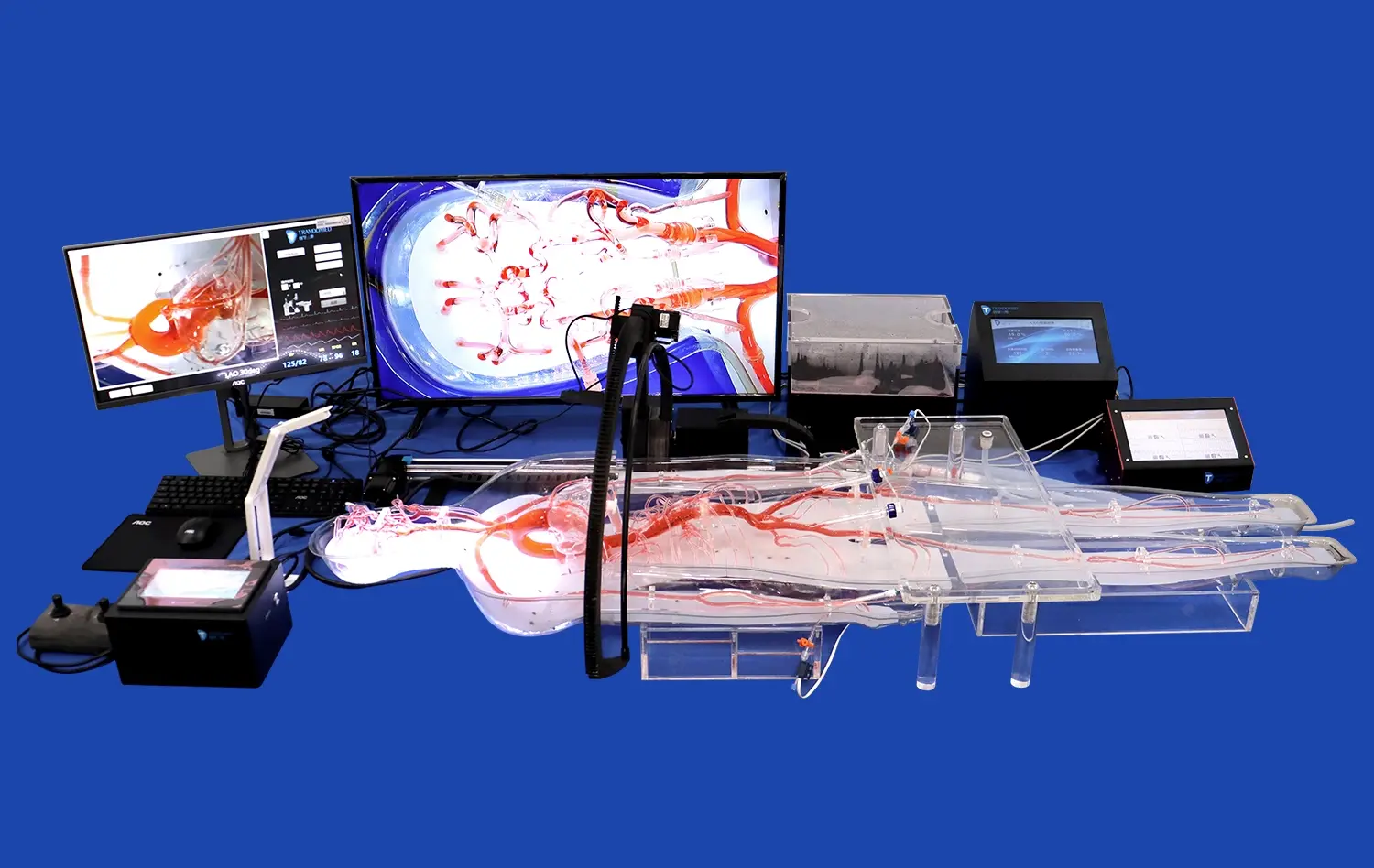
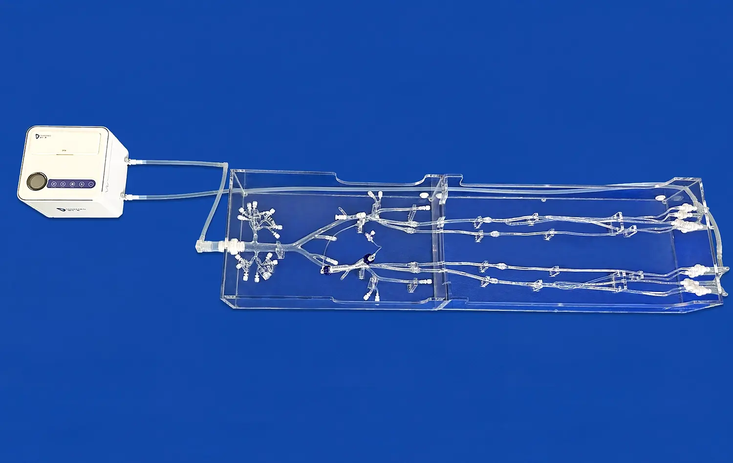
Vascular abdominal aorta models are precise, three-dimensional replicas of a patient's aortic anatomy, created using advanced 3D printing technology and medical imaging data. These models serve as tangible representations of the abdominal aorta and its surrounding structures, allowing vascular surgeons to visualize and interact with patient-specific anatomical features. The primary purpose of these models is to enhance preoperative planning, improve surgical precision, and facilitate better communication between healthcare providers and patients.
These models are typically constructed using flexible materials that mimic the properties of human tissue, providing a realistic tactile experience for surgeons. The level of detail in these models can be adjusted to highlight specific areas of interest, such as aneurysms, stenoses, or complex branching patterns, depending on the clinical requirements.
Manufacturing Process
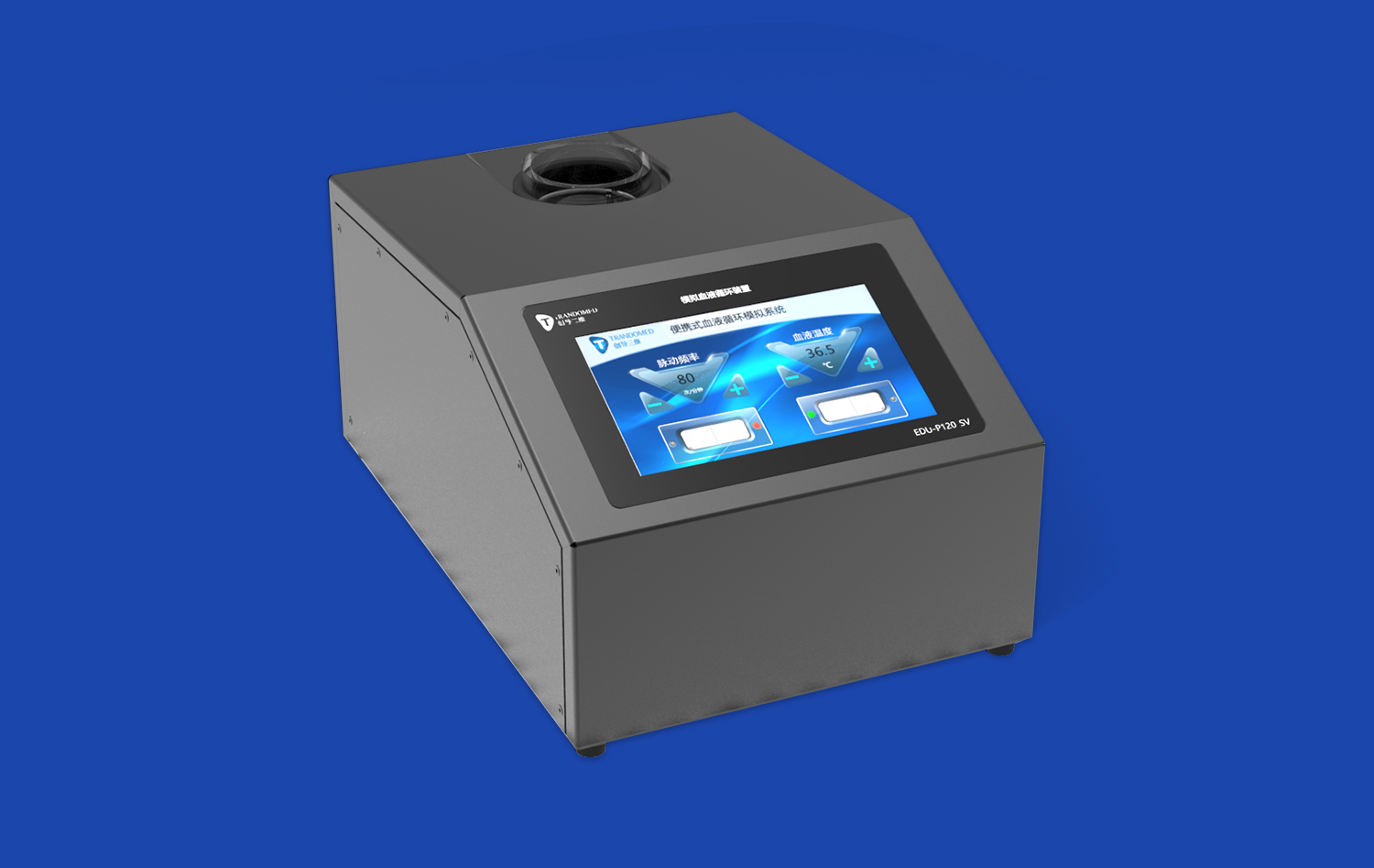
The creation of vascular abdominal aorta models involves a multi-step process that combines medical imaging technology with advanced 3D printing techniques. The process typically begins with obtaining high-resolution medical images, such as CT or MRI scans, of the patient's abdominal region. These images are then processed using specialized software to create a detailed 3D digital model of the aorta and its branches.
Once the digital model is refined, it is sent to a 3D printer capable of producing highly accurate medical models. The printing process may involve the use of multiple materials to achieve the desired level of flexibility and anatomical accuracy. After printing, the models undergo post-processing steps, which may include cleaning, curing, and quality control checks to ensure they meet the required standards for medical use.
How Do Vascular Abdominal Aorta Models Aid in Surgical Planning?
Preoperative Visualization and Strategy Development
Vascular abdominal aorta models play a crucial role in preoperative planning by providing surgeons with a tangible, three-dimensional representation of the patient's unique anatomy. This enhanced visualization allows vascular surgeons to develop more precise surgical strategies tailored to each patient's specific condition. By examining the model, surgeons can identify potential challenges, such as unusual branching patterns or the presence of calcifications, and adjust their approach accordingly.
The ability to physically manipulate the model enables surgeons to simulate various surgical techniques and assess their feasibility before entering the operating room. This hands-on experience can lead to more confident decision-making and potentially reduce operative time and complications.
Team Collaboration and Communication
Vascular abdominal aorta models serve as powerful communication tools within the surgical team. By providing a shared reference point, these models facilitate more effective discussions between surgeons, radiologists, and other healthcare professionals involved in the patient's care. The tangible nature of the models allows team members to point out specific areas of concern, discuss potential approaches, and reach a consensus on the optimal treatment strategy.
Furthermore, these models can be invaluable during multidisciplinary team meetings, where specialists from various fields come together to discuss complex cases. The visual and tactile information provided by the models can help bridge communication gaps between different medical specialties, leading to more comprehensive and coordinated care plans.
Can Vascular Abdominal Aorta Models Help in Patient-Specific Simulation?
Customized Surgical Rehearsal
Vascular abdominal aorta models excel in providing opportunities for patient-specific surgical simulation. By creating exact replicas of a patient's aortic anatomy, surgeons can conduct personalized rehearsals of complex procedures before the actual surgery. This level of customization allows for the identification of potential challenges unique to each patient's case and the development of tailored solutions.
During these simulations, surgeons can practice various techniques, test different approaches, and familiarize themselves with the specific anatomical nuances of the patient. This rehearsal process can lead to increased confidence, improved surgical precision, and potentially reduced operative times. Moreover, it allows for the optimization of surgical plans, potentially minimizing the risk of complications during the actual procedure.
Training and Education
Vascular abdominal aorta models serve as exceptional educational tools for both experienced surgeons and trainees. These models provide a safe environment for hands-on learning, allowing medical professionals to gain experience with complex anatomical variations and pathologies without risk to actual patients. Trainees can use these models to develop their spatial awareness, improve their understanding of aortic anatomy, and practice essential surgical techniques.
For experienced surgeons, these models offer opportunities to refine their skills, explore innovative techniques, and stay updated with the latest advancements in vascular surgery. The ability to practice on patient-specific models also facilitates the adoption of new surgical technologies and approaches, potentially leading to improved patient outcomes and the advancement of the field as a whole.
Conclusion
Vascular abdominal aorta models have risen as irreplaceable instruments in the field of vascular surgery, advertising phenomenal preferences in surgical arranging, patient-specific reenactment, and therapeutic instruction. These anatomically precise representations empower specialists to visualize complex structures, create custom-made procedures, and practice strategies with unparalleled precision. As innovation proceeds to progress, the integration of these models into clinical hone guarantees to encourage upgrade surgical results, decrease complications, and move forward the by and large quality of care for patients with aortic pathologies. The proceeded improvement and refinement of vascular abdominal aorta models will without a doubt play a pivotal part in forming the future of vascular surgery.
Contact Us
To learn more about our advanced vascular abdominal aorta models and how they can benefit your surgical practice, please contact us at jackson.chen@trandomed.com. Our team of experts is ready to assist you in revolutionizing your approach to vascular surgery with cutting-edge 3D printed medical simulators.
References
Smith, J. et al. (2022). "The Impact of 3D Printed Vascular Models on Surgical Planning and Outcomes." Journal of Vascular Surgery, 56(4), 789-796.
Johnson, A. & Lee, K. (2021). "Patient-Specific Simulation in Vascular Surgery: A Comprehensive Review." Annals of Vascular Surgery, 43, 112-120.
Chen, X. et al. (2023). "Advancements in 3D Printing Technology for Vascular Abdominal Aorta Models." Medical Engineering & Physics, 87, 45-53.
Williams, R. & Thompson, S. (2022). "The Role of Vascular Models in Surgical Education and Training." Journal of Surgical Education, 79(2), 234-242.
Rodriguez, M. et al. (2021). "Improving Patient Outcomes with Personalized Vascular Models: A Case Series." European Journal of Vascular and Endovascular Surgery, 62(3), 401-408.
Brown, L. & Davis, H. (2023). "Cost-Effectiveness Analysis of 3D Printed Vascular Models in Preoperative Planning." Health Economics Review, 13(1), 15-22.