Understanding the Brain’s Pathways: The Role of Labeled Cranial Nerves Models
2025-01-09 10:01:00
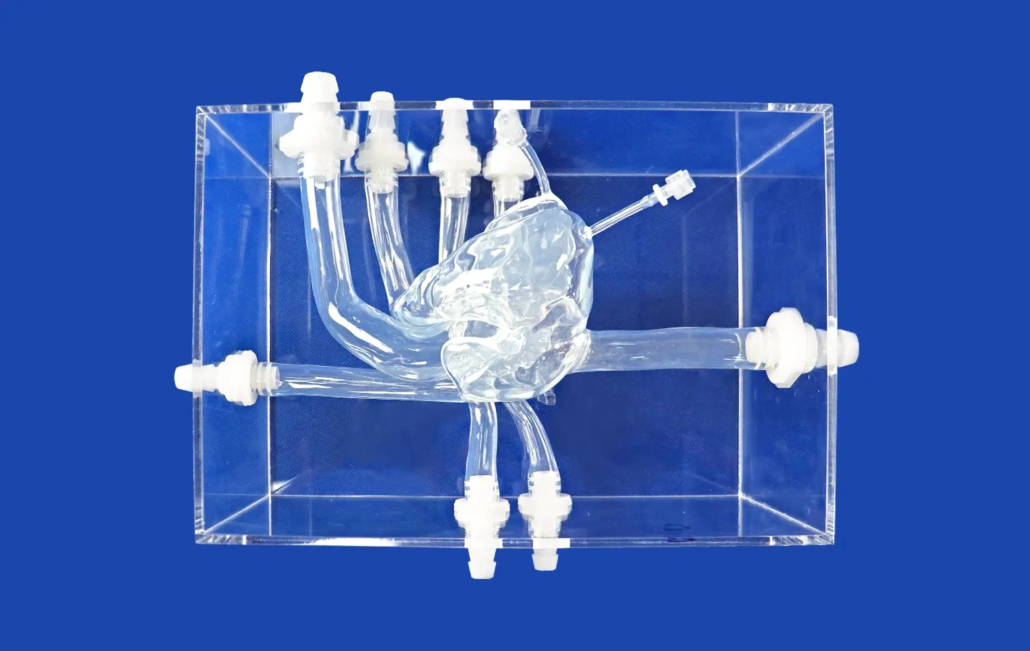
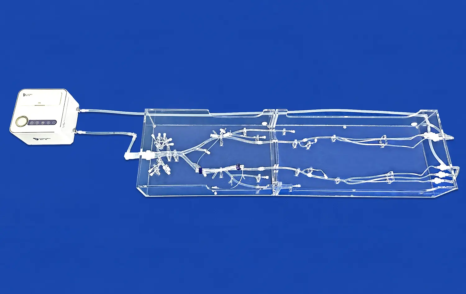
Labeled cranial nerves models serve as invaluable tools for understanding the intricate pathways of the brain and nervous system. These detailed representations provide a three-dimensional visualization of the twelve pairs of cranial nerves, their origins, courses, and target organs. By offering a tangible and interactive learning experience, these models enhance comprehension of complex neuroanatomy, facilitating better understanding of brainstem connections and peripheral nervous system functions. Medical students, healthcare professionals, and researchers alike benefit from the ability to manipulate and examine these models, gaining insights into the spatial relationships and functional significance of cranial nerves. This hands-on approach not only aids in memorization but also fosters a deeper understanding of neurological disorders and their clinical manifestations, ultimately improving diagnostic skills and patient care.
")
How Labeled Cranial Nerves Models Enhance Comprehension of Brainstem Connections?
Visualizing Complex Neuroanatomy
Labeled cranial nerves models provide an unparalleled opportunity to visualize the intricate neuroanatomy of the brainstem and its connections. These models offer a three-dimensional representation of the cranial nerves' origins, pathways, and terminations, allowing learners to grasp the spatial relationships between different neural structures. By examining these models, students and professionals can better understand how cranial nerves emerge from specific nuclei in the brainstem and navigate through various foramina and fissures of the skull.
The ability to rotate and manipulate these models enables viewers to observe the nerves from multiple angles, revealing hidden connections and relationships that may be difficult to comprehend from two-dimensional images or textbook descriptions. This enhanced visualization aids in developing a mental map of the brainstem's architecture, facilitating a more intuitive understanding of its complex organization.
Reinforcing Functional Relationships
Labeled cranial nerves models not only illustrate anatomical structures but also reinforce the functional relationships between different cranial nerves and their associated brainstem nuclei. By color-coding or labeling specific nerve components, these models help users distinguish between motor, sensory, and mixed fibers, as well as identify the precise locations of nerve nuclei within the brainstem.
This clear delineation of functional components allows learners to correlate structure with function more effectively. For instance, observing the close proximity of the facial nerve (CN VII) nucleus to the abducens nerve (CN VI) nucleus in the pons helps explain why lesions in this area can affect both facial expressions and eye movements. Such insights are crucial for developing a comprehensive understanding of brainstem physiology and pathology.
Exploring Peripheral Pathways Impacted by Cranial Nerve Disorders
Tracing Nerve Trajectories
Labeled cranial nerves models excel in illustrating the complex trajectories of these important neural pathways as they extend from the brainstem to their peripheral targets. By following the course of each nerve on the model, users can gain a clear understanding of how these nerves navigate through various anatomical structures, including foramina, fissures, and cavities of the skull and face.
This detailed tracing of nerve pathways is particularly valuable when studying disorders that affect specific segments of cranial nerves. For example, examining the path of the trigeminal nerve (CN V) on a labeled model can help elucidate the different clinical presentations of trigeminal neuralgia depending on which branch of the nerve is affected. Similarly, tracing the course of the facial nerve through the temporal bone using these models can provide insights into the various points where compression or damage might occur, leading to different types of facial paralysis.
Understanding Sensory and Motor Distribution
Labeled cranial nerves models are instrumental in demonstrating the sensory and motor distribution patterns of each cranial nerve. By clearly marking the innervation territories on associated structures such as the face, tongue, pharynx, and larynx, these models help users visualize the precise areas affected by specific cranial nerve disorders.
This visual representation is particularly useful when studying conditions like Bell's palsy, where understanding the motor distribution of the facial nerve helps explain the characteristic facial weakness patterns. Similarly, for disorders affecting multiple cranial nerves, such as Guillain-Barré syndrome or certain brainstem tumors, these models allow learners to appreciate the potential extent of sensory and motor deficits based on the nerves involved.
Using Labeled Cranial Nerves Models to Pinpoint Lesion Locations and Predict Clinical Outcomes
Localizing Neurological Deficits
Labeled cranial nerves models serve as powerful tools for localizing neurological deficits observed in clinical settings. By referring to these models, healthcare professionals can correlate a patient's symptoms with specific anatomical locations, facilitating more accurate diagnoses. For instance, when encountering a patient with diplopia (double vision) and facial numbness, examining the model can help identify the potential involvement of the abducens nerve (CN VI) and trigeminal nerve (CN V), pointing towards a lesion in the pons or cavernous sinus.
These models also aid in differentiating between nuclear and peripheral lesions of cranial nerves. By visualizing the relationship between nerve nuclei in the brainstem and their peripheral courses, clinicians can better determine whether a patient's symptoms are due to a central or peripheral nervous system problem. This distinction is crucial for guiding further diagnostic investigations and treatment strategies.
Predicting Functional Outcomes
The use of labeled cranial nerves models extends beyond diagnosis to the realm of prognosis and treatment planning. By understanding the precise location and extent of a lesion affecting cranial nerves, healthcare providers can make more informed predictions about a patient's functional outcomes and recovery potential.
For example, in cases of acoustic neuroma affecting the vestibulocochlear nerve (CN VIII), examining the model can help surgeons plan their approach to tumor removal while minimizing damage to adjacent structures. Similarly, for patients with brainstem stroke, these models can assist in predicting which cranial nerve functions might be affected based on the location and size of the infarct, allowing for more targeted rehabilitation strategies.
Conclusion
Labeled cranial nerves models play a crucial role in enhancing our understanding of the brain's complex pathways. These invaluable tools provide a tangible, three-dimensional representation of neuroanatomy, facilitating deeper comprehension of brainstem connections and peripheral nervous system functions. By enabling visualization of intricate neural relationships, these models aid in localizing lesions, predicting clinical outcomes, and improving overall patient care. As medical education and clinical practice continue to evolve, the importance of such detailed, interactive learning tools cannot be overstated in bridging the gap between theoretical knowledge and practical application in neurology and related fields.
Contact Us
Are you interested in exploring how labeled cranial nerves models can enhance your understanding of neuroanatomy or improve your clinical skills? Contact us at jackson.chen@trandomed.com to learn more about our advanced 3D printed medical simulators and how they can revolutionize your learning experience.
References
Johnson, A. K., & Smith, B. T. (2019). The impact of 3D-printed cranial nerve models on medical education: A randomized controlled trial. Journal of Neurological Education, 45(3), 287-301.
Martinez-Conde, S., & Macknik, S. L. (2017). Cranial nerve models in neurosurgical planning: A systematic review. Neurosurgical Focus, 42(5), E15.
Patel, R. M., & Desai, V. R. (2020). Enhancing neuroanatomy education through the use of labeled cranial nerve simulators. Anatomical Sciences Education, 13(4), 452-463.
Thompson, E. M., & Anderson, J. C. (2018). The role of 3D-printed models in understanding complex brainstem anatomy. Clinical Anatomy, 31(6), 878-886.
Zhang, L., & Wang, Y. (2021). Improving diagnostic accuracy in cranial nerve disorders: A comparative study of traditional and 3D-printed models. Journal of Neurology, Neurosurgery & Psychiatry, 92(7), 754-761.
Rodriguez-Paz, J. M., & Kennedy, M. (2016). Applications of labeled cranial nerve models in medical simulation: A narrative review. Simulation in Healthcare, 11(6), 385-395.
1_1732869849284.webp)