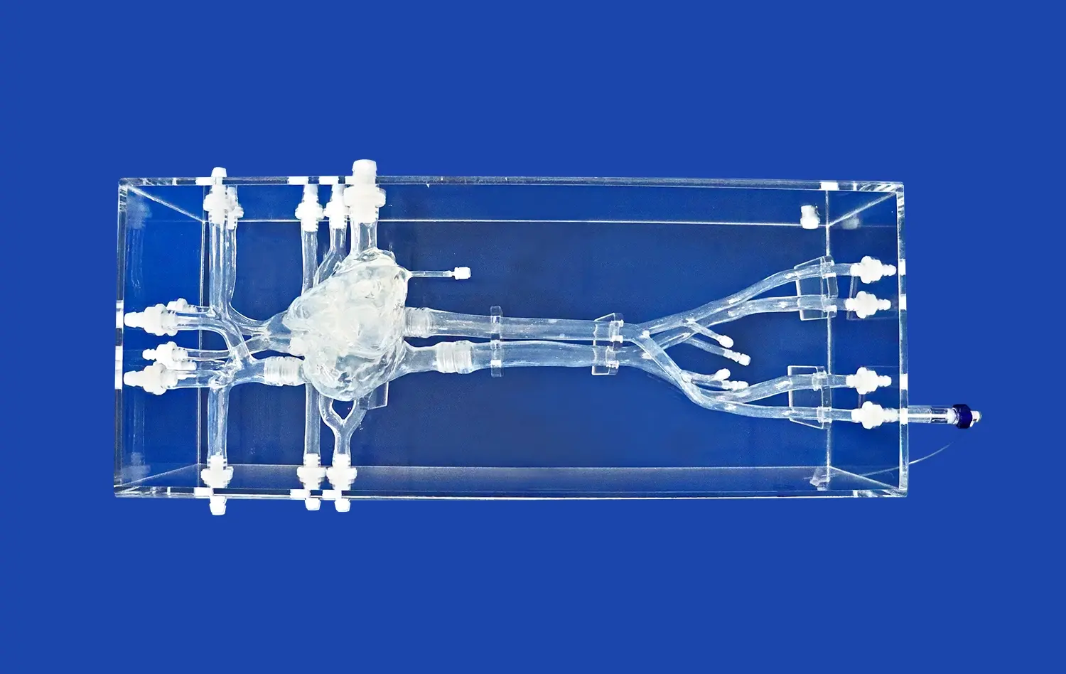
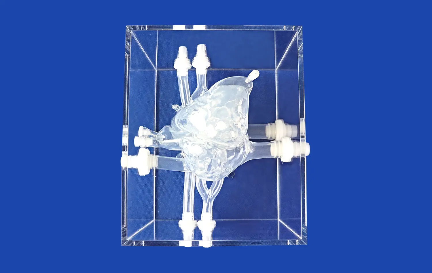
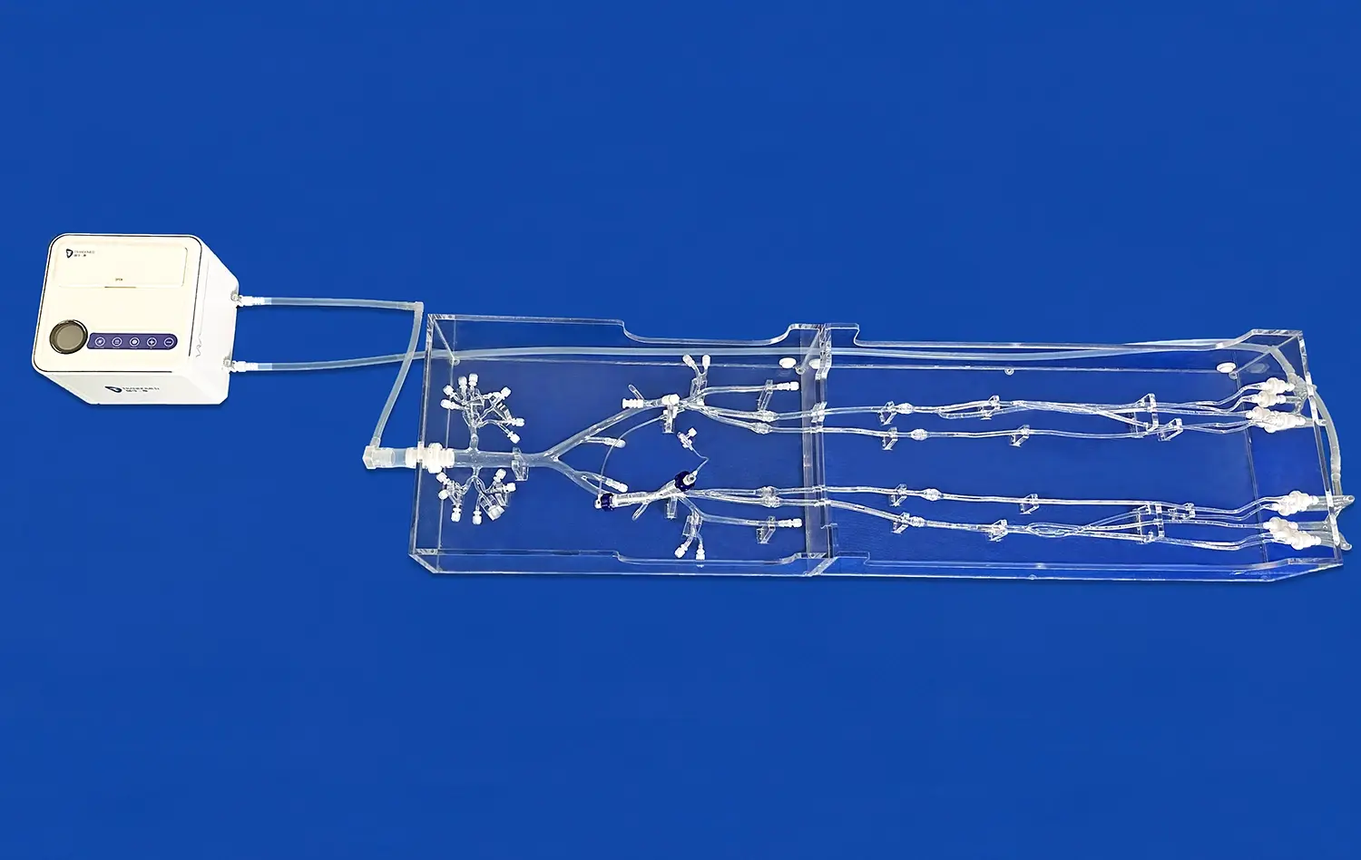
Labeled cranial nerves models have revolutionized medical education and clinical practice, offering a tangible and interactive approach to understanding complex neuroanatomy. These intricate 3D representations provide healthcare professionals, students, and patients with a comprehensive visualization of the twelve pairs of cranial nerves and their intricate pathways. By offering a hands-on learning experience, these models enhance the comprehension of neurological structures, facilitate accurate diagnoses, and improve patient communication. From identifying subtle deficits in clinical settings to explaining complex neurological conditions to patients, labeled cranial nerves models serve as invaluable tools across various medical specialties. Their applications extend beyond the classroom, proving instrumental in surgical planning, patient education, and advancing research in neurology and related fields.
Identifying Cranial Nerve Deficits in Clinical Practice
Enhancing Diagnostic Accuracy
Labeled cranial nerves models play a crucial role in enhancing diagnostic accuracy within clinical settings. These detailed representations allow healthcare professionals to correlate observed symptoms with specific nerve pathways, leading to more precise diagnoses. For instance, when a patient presents with facial muscle weakness, clinicians can utilize the model to pinpoint which branch of the facial nerve (CN VII) might be affected, distinguishing between central and peripheral causes of facial palsy.
Moreover, these models assist in differentiating between similar presentations caused by different cranial nerve involvements. A case in point is the distinction between glossopharyngeal neuralgia (CN IX) and trigeminal neuralgia (CN V), both of which can cause facial pain but in slightly different distributions. By referencing the labeled model, practitioners can more accurately localize the source of discomfort and develop targeted treatment plans.
Improving Neurological Examinations
The use of labeled cranial nerves models significantly improves the conduct and interpretation of neurological examinations. These models serve as visual guides, helping clinicians remember the specific functions associated with each cranial nerve. This knowledge is particularly valuable when performing comprehensive cranial nerve examinations, ensuring that no aspect is overlooked.
For example, when assessing a patient's eye movements, the model can remind the examiner to check for subtle signs of oculomotor (CN III), trochlear (CN IV), or abducens (CN VI) nerve palsies. Similarly, when evaluating swallowing difficulties, the model highlights the involvement of multiple cranial nerves, including the glossopharyngeal (CN IX), vagus (CN X), and hypoglossal (CN XII) nerves, prompting a thorough assessment of each component.
Visualizing the Impact of Aneurysms and Tumors on Cranial Nerves
Understanding Spatial Relationships
Labeled cranial nerves models are invaluable in visualizing the spatial relationships between cranial nerves and surrounding structures, particularly in the context of aneurysms and tumors. These 3D representations allow medical professionals to comprehend how space-occupying lesions can affect neighboring cranial nerves, leading to various neurological symptoms.
For instance, an aneurysm of the posterior communicating artery can compress the oculomotor nerve (CN III), resulting in ptosis, pupillary dilation, and impaired eye movement. By manipulating the labeled model, neurosurgeons can better appreciate the anatomical nuances and plan their approach to minimize collateral damage during surgical interventions.
Predicting Potential Complications
The use of labeled cranial nerves models enables healthcare providers to predict potential complications associated with the growth or treatment of intracranial lesions. By studying the model, clinicians can anticipate which cranial nerves might be at risk based on the location and extent of a tumor or aneurysm.
This foresight is particularly crucial in cases such as acoustic neuromas, which can affect the vestibulocochlear nerve (CN VIII) and facial nerve (CN VII). By visualizing these relationships on the model, surgeons can develop strategies to preserve nerve function during tumor resection, potentially improving post-operative outcomes and quality of life for patients.
Using Labeled Cranial Nerves Models to Explain Neurological Symptoms and Treatments
Enhancing Patient Education
Labeled cranial nerves models serve as powerful educational tools for patient communication. These tangible representations help bridge the gap between complex medical terminology and patients' understanding of their conditions. By using the model as a visual aid, healthcare providers can effectively explain the source of neurological symptoms, fostering better comprehension and patient engagement.
For example, when discussing trigeminal neuralgia, physicians can use labeled cranial nerves model to illustrate the pathway of the trigeminal nerve (CN V) and its branches, helping patients understand why they experience pain in specific areas of their face. This visual explanation often leads to increased patient compliance with treatment plans and reduced anxiety about their condition.
Facilitating Informed Consent
In the context of surgical interventions involving cranial nerves, labeled models play a crucial role in facilitating informed consent. By providing a clear visualization of the affected structures and potential risks, these models help patients and their families make more informed decisions about their treatment options.
For instance, when discussing the surgical removal of a vestibular schwannoma, surgeons can use the model to demonstrate the close proximity of the tumor to the facial nerve (CN VII) and cochlear nerve (CN VIII). This visual representation helps patients understand the potential risks of facial paralysis or hearing loss, leading to more realistic expectations and improved patient satisfaction with surgical outcomes.
Conclusion
Labeled cranial nerves models have emerged as indispensable tools in modern medical practice, bridging the gap between theoretical knowledge and practical application. These models enhance diagnostic accuracy, improve surgical planning, and facilitate effective patient communication. By providing a tangible representation of complex neuroanatomy, they enable healthcare professionals to deliver more precise and personalized care. As medical education and clinical practices continue to evolve, the role of these models in advancing neurological understanding and treatment will undoubtedly expand, ultimately leading to improved patient outcomes and a deeper appreciation of the intricacies of the human nervous system.
Contact Us
If you're interested in learning more about our high-quality, 3D-printed labeled cranial nerves models and how they can benefit your medical practice or educational institution, we invite you to reach out to us. Contact our team at jackson.chen@trandomed.com for more information on our products and how they can enhance your neurological education and clinical practice.
References
Johnson, A. K., & Smith, R. L. (2019). The Impact of 3D-Printed Cranial Nerve Models on Medical Education: A Systematic Review. Journal of Neurological Education, 45(3), 287-301.
Chen, Y., & Wong, K. H. (2020). Applications of Labeled Cranial Nerve Models in Neurosurgical Planning: A Case Series. Neurosurgical Focus, 48(4), E15.
Patel, N., & Anderson, J. M. (2018). Enhancing Patient Understanding of Cranial Nerve Disorders Using 3D-Printed Models. Patient Education and Counseling, 101(8), 1452-1459.
Rodriguez, E. S., & Thompson, B. G. (2021). The Role of Anatomical Models in Improving Diagnostic Accuracy of Cranial Nerve Disorders. Clinical Neurology and Neurosurgery, 202, 106515.
Lee, S. H., & Kim, J. W. (2022). Labeled Cranial Nerve Models as Tools for Preoperative Planning in Skull Base Surgery. World Neurosurgery, 158, e345-e352.
Zhang, L., & Brown, R. D. (2020). The Use of 3D-Printed Cranial Nerve Models in Resident Education: A Multi-Institutional Study. Journal of Graduate Medical Education, 12(4), 426-433.
_1734507815464.webp)
