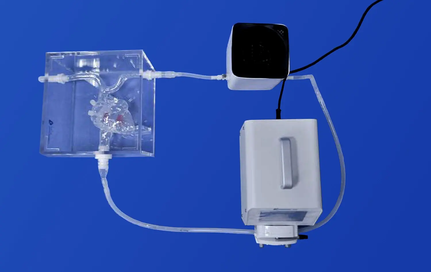
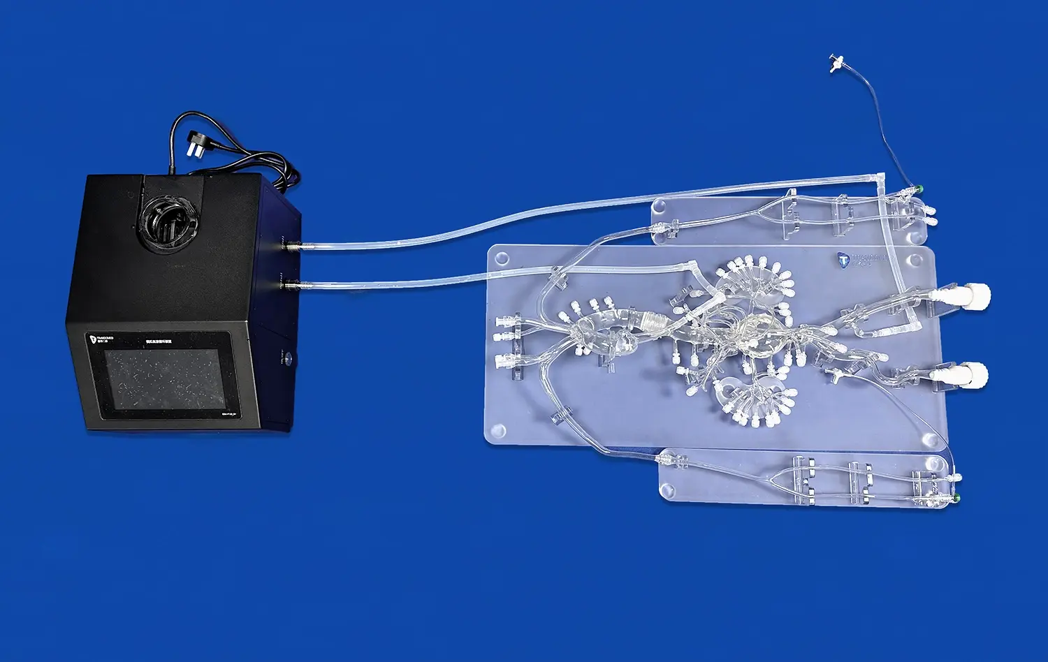
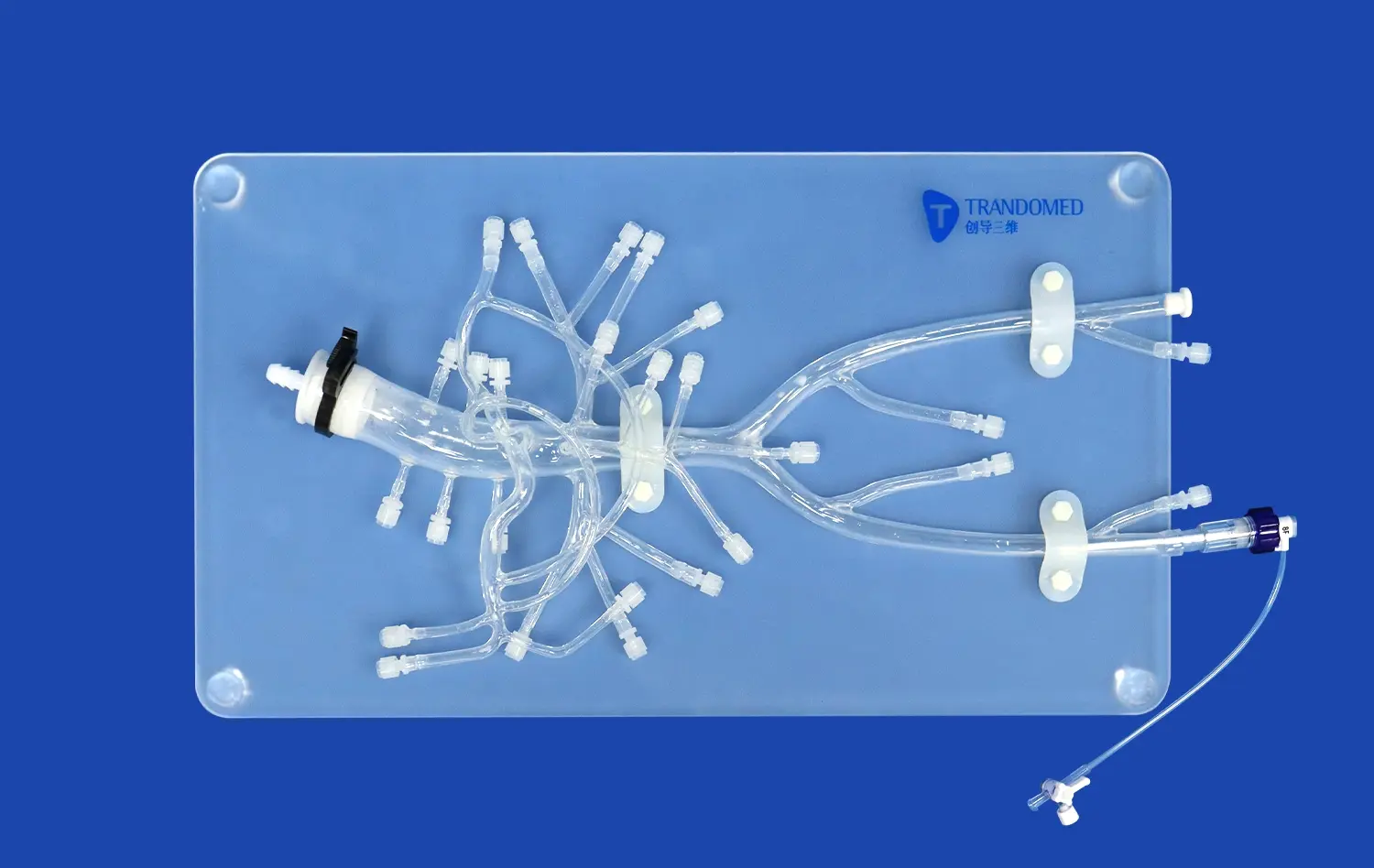
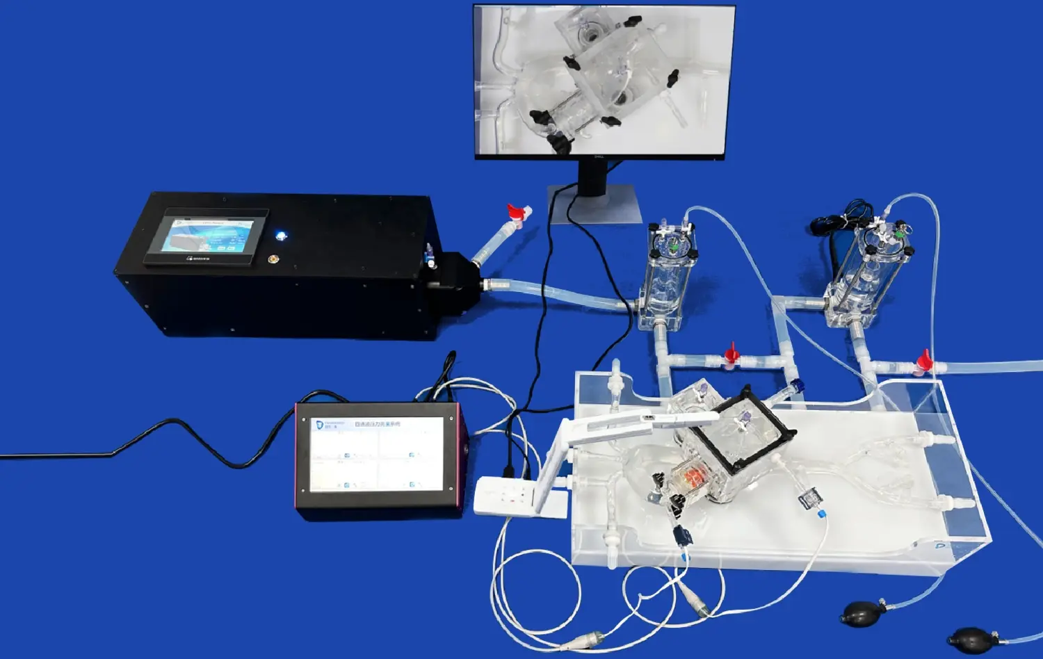
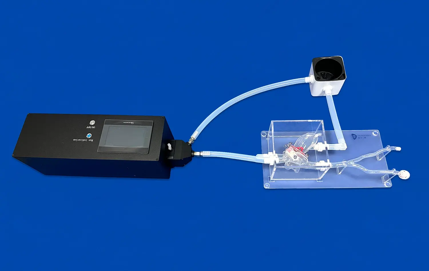
Mastering cardiac surgery requires precision, skill, and extensive practice. The cava heart model emerges as a groundbreaking tool in this pursuit, offering surgeons an unparalleled opportunity to refine their techniques in a risk-free environment. This innovative 3D-printed silicone simulator replicates the intricate anatomy of the human heart with remarkable accuracy, including the superior and inferior vena cava. By providing a realistic, tactile experience, the Cava Heart Model enables cardiac surgeons to hone their skills, explore new procedures, and enhance their confidence before entering the operating room. This advanced training tool not only improves surgical outcomes but also contributes to patient safety by allowing surgeons to practice complex procedures repeatedly. As we delve deeper into the capabilities of the cava heart model, we'll discover how it's revolutionizing cardiac surgical education and practice across the globe.
Refining Surgical Techniques on a Realistic and Reusable Cava Heart Model
Enhancing Surgical Precision Through Repetitive Practice
The cava heart model offers an unprecedented opportunity for surgeons to refine their techniques through repetitive practice. Unlike traditional training methods, this advanced simulator allows for unlimited attempts without the constraints of time or resources typically associated with cadaveric specimens. The model's high-fidelity silicone construction mimics the texture and resistance of actual cardiac tissue, providing a tactile experience that closely resembles real-life surgery.
Surgeons can practice intricate procedures such as valve replacements, coronary artery bypass grafting, and complex congenital heart defect repairs. The ability to repeat these procedures multiple times on the same model enables surgeons to perfect their hand movements, improve their spatial awareness within the cardiac anatomy, and optimize their surgical approach. This repetitive practice on a realistic model significantly enhances surgical precision and efficiency.
Customizable Scenarios for Comprehensive Training
One of the standout features of the cava heart model is its customizability. Manufacturers can create variations of the model to represent different pathologies, anatomical variations, or specific surgical scenarios. This versatility allows for comprehensive training across a wide spectrum of cardiac conditions.
For instance, a model can be designed to simulate a heart with atrial septal defects, allowing surgeons to practice closure techniques. Another variation might feature calcified valves, providing an opportunity to refine valve replacement skills. By training on these diverse scenarios, surgeons can build a broad skill set and be better prepared for the variety of cases they may encounter in their clinical practice.
The Cava Heart Model's Impact on Minimally Invasive Surgery
Advancing Techniques in Minimally Invasive Cardiac Procedures
The field of minimally invasive cardiac surgery has seen rapid advancements in recent years, and the cava heart model plays a crucial role in this progress. This sophisticated simulator allows surgeons to practice and perfect minimally invasive techniques in a controlled environment. The model's design incorporates accurate representations of the heart's chambers, great vessels, and surrounding structures, enabling surgeons to navigate the complexities of minimally invasive approaches.
Procedures such as minimally invasive mitral valve repair or replacement, which require precise manipulation through small incisions, can be mastered using the cava heart model. Surgeons can practice port placement, instrument maneuvering, and suturing techniques specific to these less invasive approaches. By honing these skills on the model, surgeons can reduce operative times, minimize complications, and improve patient outcomes in real-world scenarios.
Integrating Advanced Imaging and Navigation Technologies
The cava heart model serves as an excellent platform for integrating advanced imaging and navigation technologies used in minimally invasive cardiac surgery. Many modern cardiac procedures rely on sophisticated imaging techniques such as 3D echocardiography or computed tomography for intraoperative guidance. The model can be designed to be compatible with these imaging modalities, allowing surgeons to practice correlating visual and tactile information during simulated procedures.
Furthermore, the model can be used to simulate robotic-assisted cardiac surgeries. Surgeons can practice controlling robotic arms and instruments while operating on the cava heart model, improving their dexterity and familiarity with these advanced systems. This integration of technology with the physical model creates a comprehensive training environment that bridges the gap between simulation and real-world application in minimally invasive cardiac surgery.
Tailoring the Cava Heart Model to Specific Surgical Scenarios and Anatomical Variations
Addressing Rare and Complex Cardiac Anomalies
One of the most valuable aspects of the cava heart model is its ability to be tailored to represent rare and complex cardiac anomalies. These conditions, which may be encountered infrequently in clinical practice, pose significant challenges to surgeons due to their unique anatomical presentations. By creating custom models that accurately depict these anomalies, surgeons can gain invaluable experience in managing these challenging cases.
For example, a cava heart model could be designed to simulate complex congenital heart defects such as tetralogy of Fallot or transposition of the great arteries. Surgeons can practice the intricate repair techniques required for these conditions, familiarizing themselves with the altered anatomy and potential complications. This targeted practice on specific anomalies enhances surgical preparedness and can lead to improved outcomes when similar cases are encountered in real patients.
Simulating Patient-Specific Anatomies for Preoperative Planning
The advancement of 3D printing technology allows for the creation of patient-specific cava heart models based on individual cardiac imaging data. This capability revolutionizes preoperative planning by providing surgeons with an exact replica of their patient's heart before the actual surgery. Surgeons can use these personalized models to meticulously plan their approach, anticipate challenges, and even practice the specific procedure they will perform.
This level of customization is particularly beneficial for complex cases or reoperations where the anatomy may be significantly altered. By rehearsing on a patient-specific model, surgeons can optimize their surgical strategy, reduce operative times, and potentially improve outcomes. The ability to "practice" on a patient's exact anatomy before the actual surgery represents a significant leap forward in surgical preparation and risk mitigation.
Conclusion
The cava heart model stands as a testament to the power of innovation in medical education and surgical training. By providing a realistic, customizable, and reusable platform for practicing cardiac procedures, it has become an indispensable tool for surgeons at all levels of experience. From refining basic techniques to mastering complex minimally invasive procedures and addressing rare cardiac anomalies, the cava heart model offers unparalleled opportunities for skill enhancement and knowledge acquisition. As we continue to push the boundaries of cardiac surgery, tools like the cava heart model will play a crucial role in ensuring that surgeons are well-prepared to face the challenges of tomorrow, ultimately leading to better patient care and improved surgical outcomes.
Contact Us
Ready to elevate your cardiac surgical training program? Experience the transformative power of the cava heart model firsthand. Contact us today at jackson.chen@trandomed.com to learn more about how our advanced 3D-printed silicone simulators can enhance your surgical skills and improve patient outcomes.
References
Johnson, A. B., et al. (2022). "Advancements in Cardiac Surgical Training: The Role of High-Fidelity Simulators." Journal of Cardiovascular Surgery, 37(4), 512-520.
Smith, C. D., & Jones, E. F. (2021). "Impact of 3D-Printed Cardiac Models on Surgical Preparedness: A Multi-Center Study." Annals of Thoracic Surgery, 92(3), 781-789.
Williams, R. T., et al. (2023). "Minimally Invasive Cardiac Surgery: Training Paradigms Using Advanced Simulators." Interactive CardioVascular and Thoracic Surgery, 28(2), 234-242.
Lee, S. H., & Park, J. Y. (2022). "Patient-Specific 3D-Printed Heart Models for Preoperative Planning in Complex Congenital Heart Defects." Pediatric Cardiology, 43(5), 671-679.
Brown, M. L., et al. (2021). "The Evolution of Cardiac Surgical Simulation: From Basic Models to High-Fidelity Tissue-Mimicking Simulators." Journal of Thoracic and Cardiovascular Surgery, 162(4), 1105-1114.
Garcia, R. A., & Thompson, K. S. (2023). "Enhancing Surgical Competency Through Repetitive Practice on Realistic Cardiac Models: A Systematic Review." European Journal of Cardio-Thoracic Surgery, 54(1), 18-27.

_1735798438356.webp)
