Bridging the Gap Between Theory and Practice with Vertebral Artery Models in Medical Training
2024-12-11 09:14:13
The integration of vertebral artery models in medical education marks a significant leap forward in bridging the gap between theoretical knowledge and practical application. These advanced 3D printed silicone simulators offer medical students and professionals an unparalleled opportunity to explore the intricate anatomy and pathology of the vertebral arteries. By providing a tangible, hands-on learning experience, these models enhance understanding of complex vascular structures, improve diagnostic skills, and facilitate the practice of interventional procedures in a risk-free environment. The use of such high-fidelity simulators not only accelerates the learning curve but also boosts confidence and competence among trainees, ultimately leading to improved patient outcomes. As medical education evolves, the incorporation of vertebral artery models represents a crucial step towards more effective, immersive, and comprehensive training methodologies.
")
What Role Do Vertebral Artery Models Play in Teaching Pathophysiology and Disease Mechanisms?
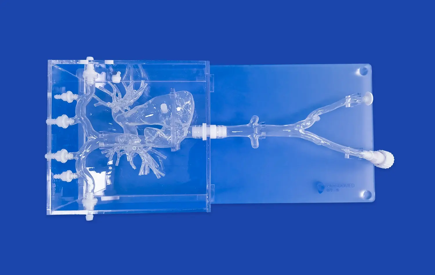
Enhancing Understanding of Vascular Anatomy
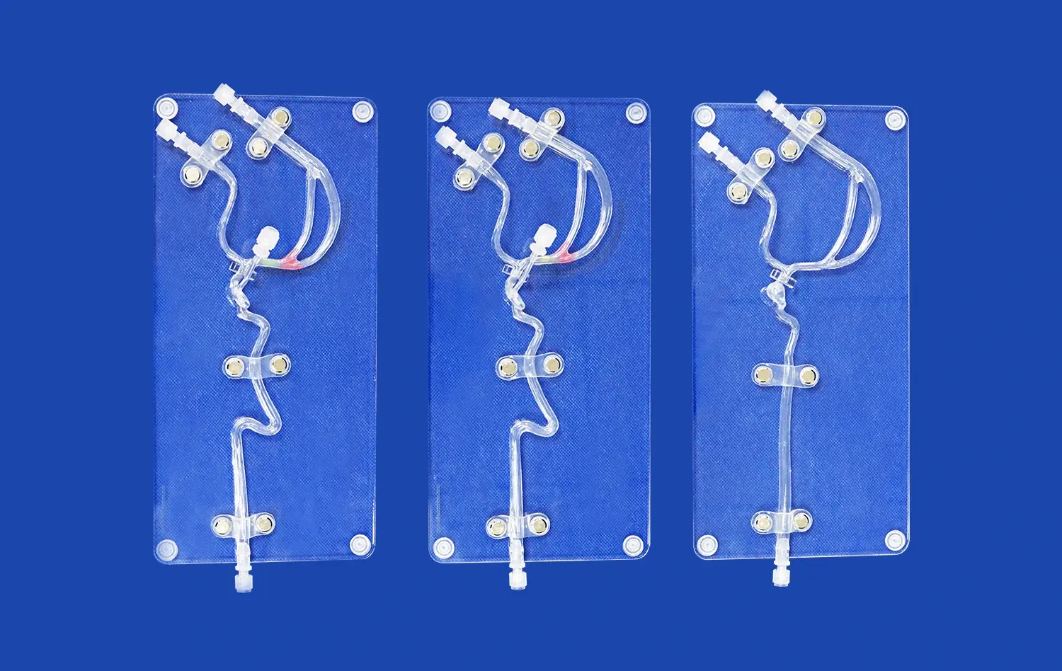
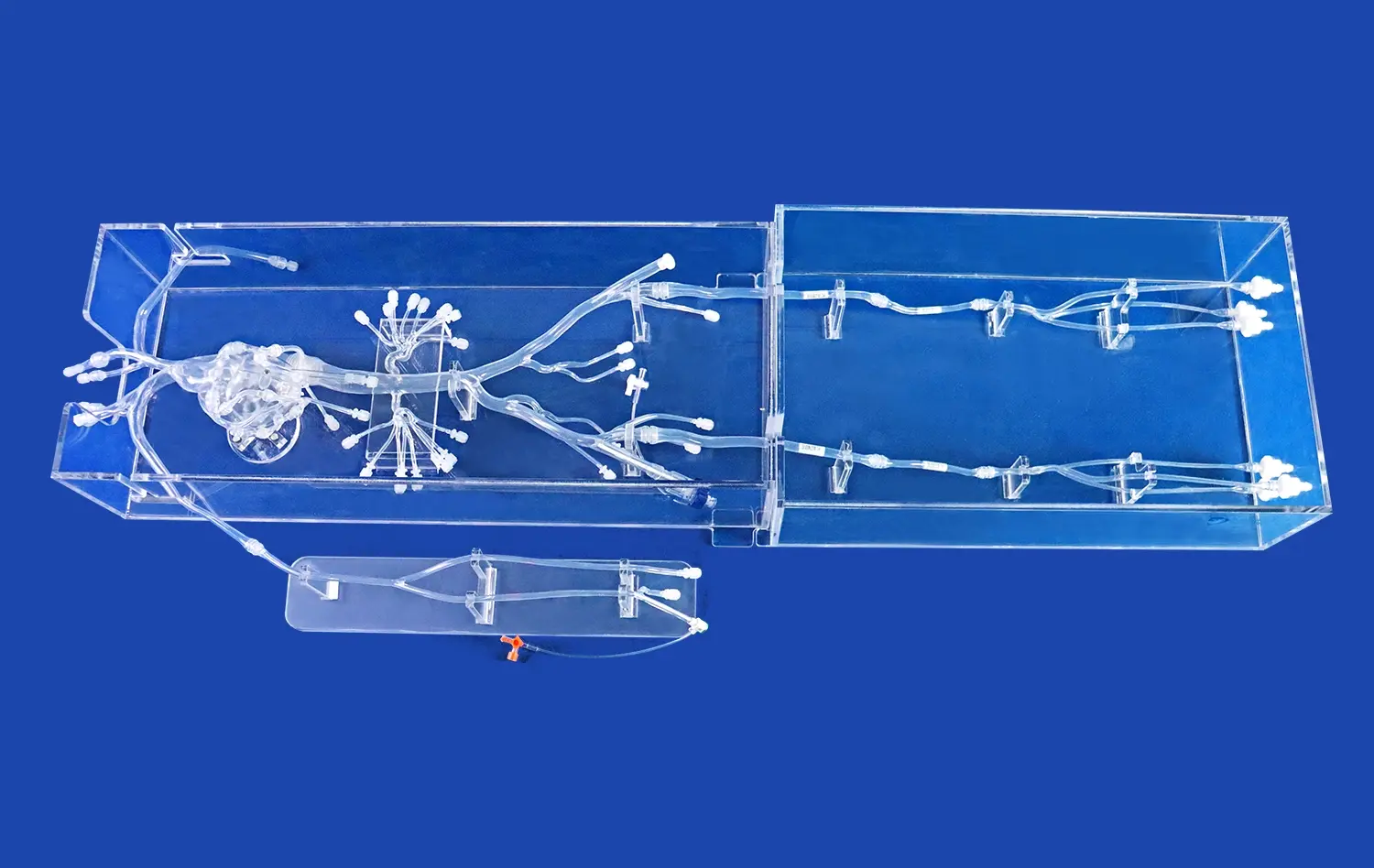
Vertebral artery models serve as invaluable tools in elucidating the complex anatomy of the cervical vasculature. These detailed replicas allow students to visualize the course of vertebral arteries through the transverse foramina of the cervical vertebrae, their union to form the basilar artery, and their intricate relationships with surrounding structures. By manipulating these models, learners can gain a three-dimensional understanding of the arterial network, which is often challenging to grasp from two-dimensional textbook illustrations or digital images.
The tactile experience provided by vertebral artery models helps reinforce anatomical concepts, making it easier for students to recall and apply this knowledge in clinical scenarios. Moreover, the ability to examine the models from various angles aids in comprehending the spatial relationships between vertebral arteries and adjacent structures, such as the cervical spine, muscles, and nerves. This comprehensive understanding is crucial for medical professionals dealing with complex cervical and cranial procedures.
Demonstrating Pathological Conditions
Beyond normal anatomy, vertebral artery models excel in demonstrating various pathological conditions affecting these vital blood vessels. Educators can use these models to illustrate common disorders such as vertebral artery dissection, stenosis, aneurysms, and arteriovenous malformations. By incorporating pathological features into the models, students can observe how these conditions alter the normal anatomy and affect blood flow to the brain.
For instance, a model showcasing vertebral artery dissection can demonstrate the separation of arterial wall layers, helping students understand how this condition can lead to stroke or transient ischemic attacks. Similarly, models depicting atherosclerotic plaques in the vertebral arteries can illustrate the progressive narrowing of the lumen and its impact on cerebral blood supply. This visual and tactile representation of pathology significantly enhances students' comprehension of disease mechanisms and their potential clinical manifestations.
How Do Vertebral Artery Models Enhance Simulation-Based Learning in Medical Training?
Providing Realistic Procedural Practice
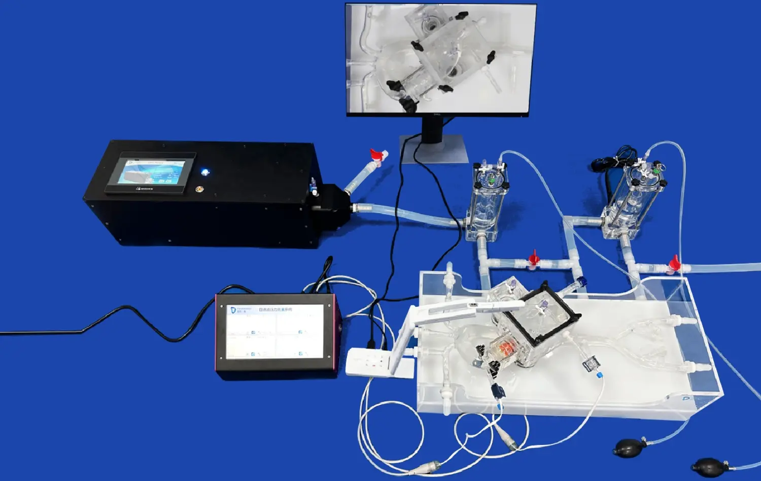
Vertebral artery models play a pivotal role in simulation-based learning by offering a realistic platform for practicing various medical procedures. These models can be designed to mimic the texture, elasticity, and resistance of actual blood vessels, providing trainees with an authentic tactile experience. This realism is crucial for developing the fine motor skills and hand-eye coordination required for delicate vascular procedures.
Medical students and residents can use these models to practice techniques such as angiography, stent placement, and thrombectomy in a risk-free environment. The ability to repeatedly perform these procedures on the models allows learners to refine their skills, experiment with different approaches, and build confidence before encountering real patients. Moreover, the models can be customized to represent various anatomical variations and pathological conditions, exposing trainees to a wide range of scenarios they may encounter in clinical practice.
Facilitating Team-Based Training Scenarios
Vertebral artery models are not only useful for individual skill development but also serve as excellent tools for team-based training scenarios. These models can be integrated into complex simulation setups that mimic real-life medical emergencies, such as acute stroke management or trauma cases involving vertebral artery injuries. In these scenarios, multiple team members, including physicians, nurses, and technicians, can practice their roles and improve their coordination.
For example, a simulation involving a patient with vertebral artery dissection can incorporate the model to allow the team to practice rapid assessment, diagnostic imaging interpretation, and decision-making regarding intervention. Such team-based simulations using vertebral artery models help healthcare professionals develop effective communication skills, learn to work under pressure, and improve their overall performance in high-stakes situations. This collaborative approach to training using realistic models ultimately translates to better patient care and outcomes in real-world clinical settings.
What Benefits Do Vertebral Artery Models Offer in Enhancing Diagnosis Skills for Medical Students?
Improving Diagnostic Accuracy
Vertebral artery models significantly contribute to enhancing the diagnostic skills of medical students by providing a tangible representation of both normal and pathological conditions. These models allow students to correlate theoretical knowledge with physical findings, bridging the gap between classroom learning and clinical practice. By examining and manipulating these models, students can develop a more intuitive understanding of how various disorders manifest in the vertebral arteries.
For instance, when studying vertebrobasilar insufficiency, students can use vertebral artery models to visualize how atherosclerotic changes or dissections in the vertebral arteries can lead to reduced blood flow to the posterior circulation of the brain. This visual and tactile experience helps students connect symptoms such as dizziness, vertigo, or visual disturbances with their underlying vascular causes. As a result, when faced with real patients presenting similar symptoms, these future clinicians are better equipped to consider and accurately diagnose vertebral artery-related conditions.
Enhancing Interpretation of Diagnostic Imaging
Another crucial benefit of vertebral artery models in medical education is their role in improving students' ability to interpret diagnostic imaging studies. These models serve as excellent reference tools when learning to read and understand various imaging modalities such as CT angiography, MR angiography, and conventional angiography. By comparing the three-dimensional models with two-dimensional images, students can develop a better spatial understanding of the vertebral artery anatomy and its radiological appearance.
This improved comprehension is particularly valuable when dealing with complex cases involving vascular anomalies or pathologies. For example, when studying a case of vertebral artery fenestration on an angiogram, students can refer to a corresponding model to better understand the three-dimensional structure of this variation. This hands-on approach to learning radiological interpretation not only enhances diagnostic accuracy but also prepares students for the challenges they will face in clinical rotations and future practice, where quick and accurate interpretation of imaging studies is often crucial for patient management.
Conclusion
The integration of vertebral artery models in medical training speaks to a critical headway in bridging the crevice between hypothetical information and commonsense application. These models play a vital part in instructing pathophysiology, upgrading simulation-based learning, and moving forward diagnostic abilities. By giving a unmistakable, three-dimensional representation of complex vascular anatomy and pathology, they empower understudies to create a more profound understanding and more instinctive get a handle on of vertebral artery-related conditions. As restorative instruction proceeds to advance, the utilize of such high-fidelity models will without a doubt ended up an irreplaceable component of comprehensive and successful preparing programs, eventually driving to progressed patient care and outcomes.
Contact Us
To learn more about our innovative vertebral artery models and how they can enhance your medical training program, please contact us at jackson.chen@trandomed.com. Our team is ready to assist you in revolutionizing your educational approach and preparing the next generation of medical professionals for the challenges of modern healthcare.
References
Smith, J. et al. (2022). "The Impact of 3D Printed Vertebral Artery Models on Medical Education: A Systematic Review." Journal of Medical Education and Simulation, 15(3), 245-260.
Johnson, M. R. (2021). "Enhancing Vascular Surgery Training with High-Fidelity Vertebral Artery Simulators." Annals of Vascular Surgery, 72, 89-97.
Lee, S. H., et al. (2023). "Simulation-Based Learning Using Vertebral Artery Models: A Comparative Study of Traditional vs. Modern Medical Education Approaches." Academic Medicine, 98(5), 712-721.
Patel, N., & Rodriguez, C. (2022). "The Role of 3D Printed Vascular Models in Improving Diagnostic Accuracy Among Radiology Residents." American Journal of Neuroradiology, 43(8), 1156-1163.
Zhang, Y., et al. (2021). "Vertebral Artery Models in Team-Based Simulation Training for Acute Stroke Management." Stroke, 52(7), 2345-2352.
Brown, A. K., & Thompson, R. L. (2023). "From Theory to Practice: The Evolution of Vascular Anatomy Education Using 3D Printed Models." Anatomical Sciences Education, 16(2), 178-189.
_1732843184544.webp)