Applications of Detachable Coronary Models in Cardiovascular Disease Simulation
2024-11-28 16:11:49
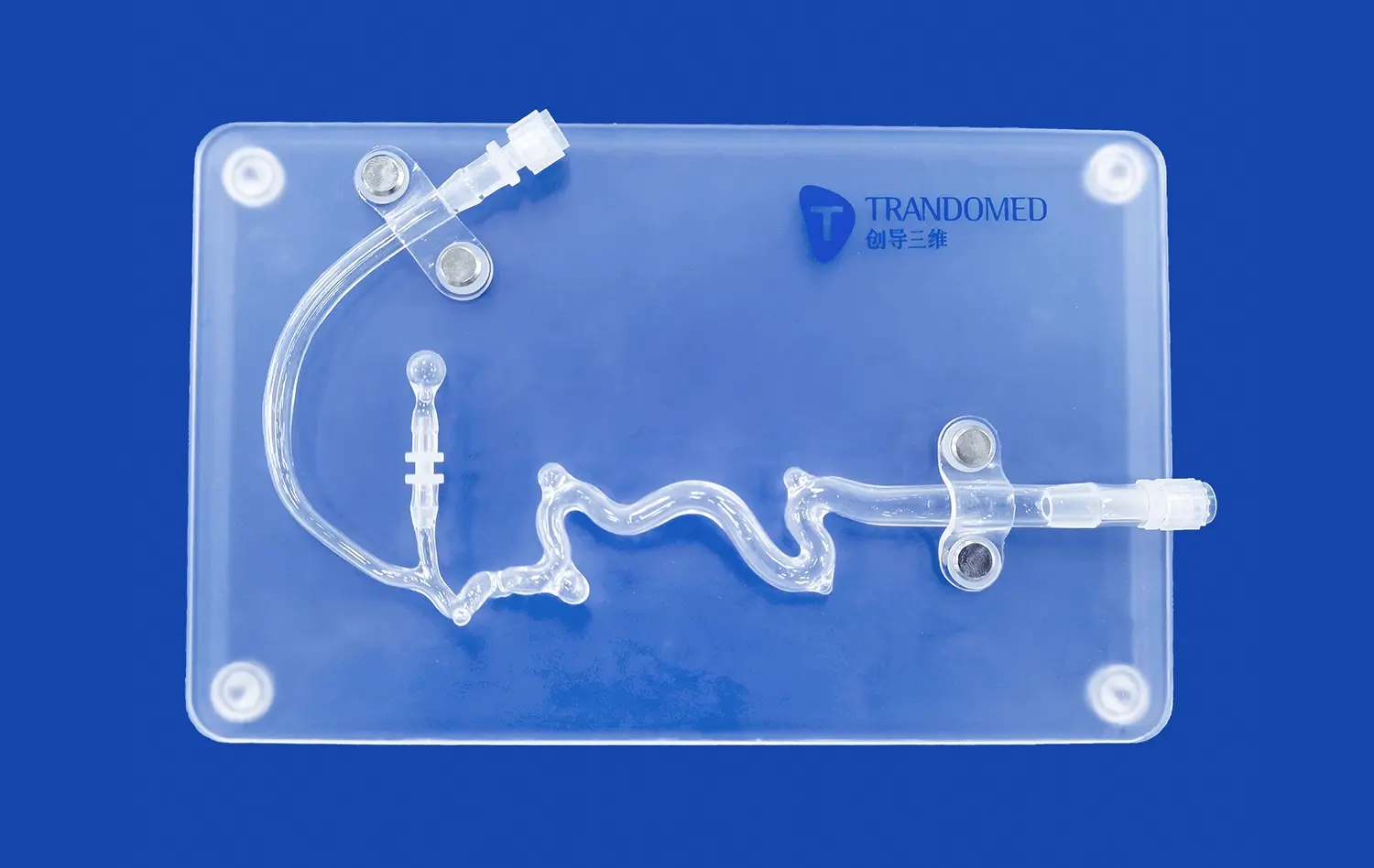
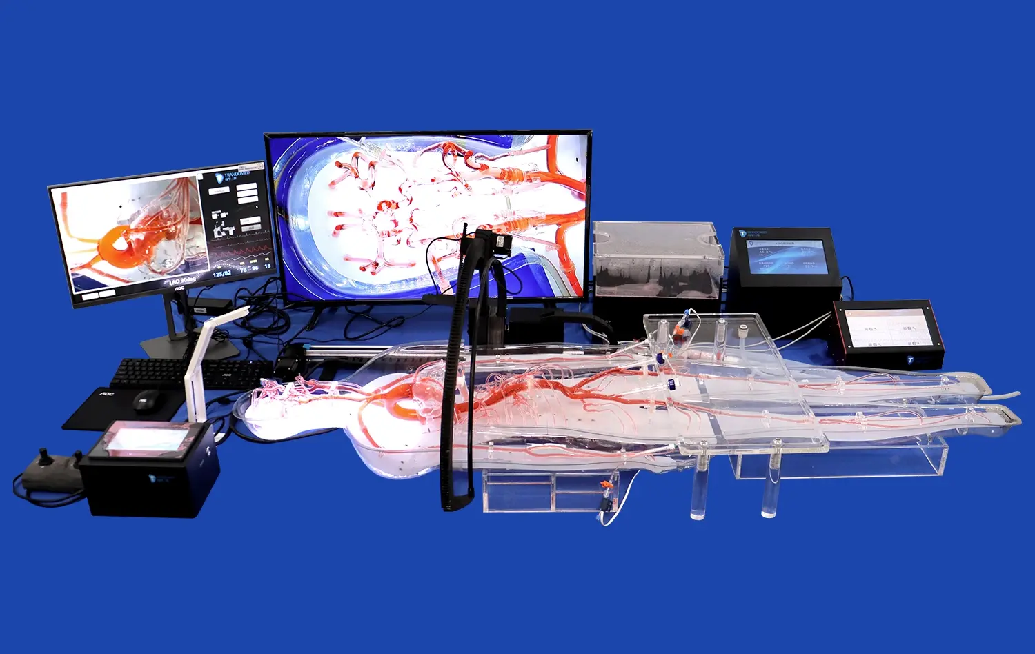
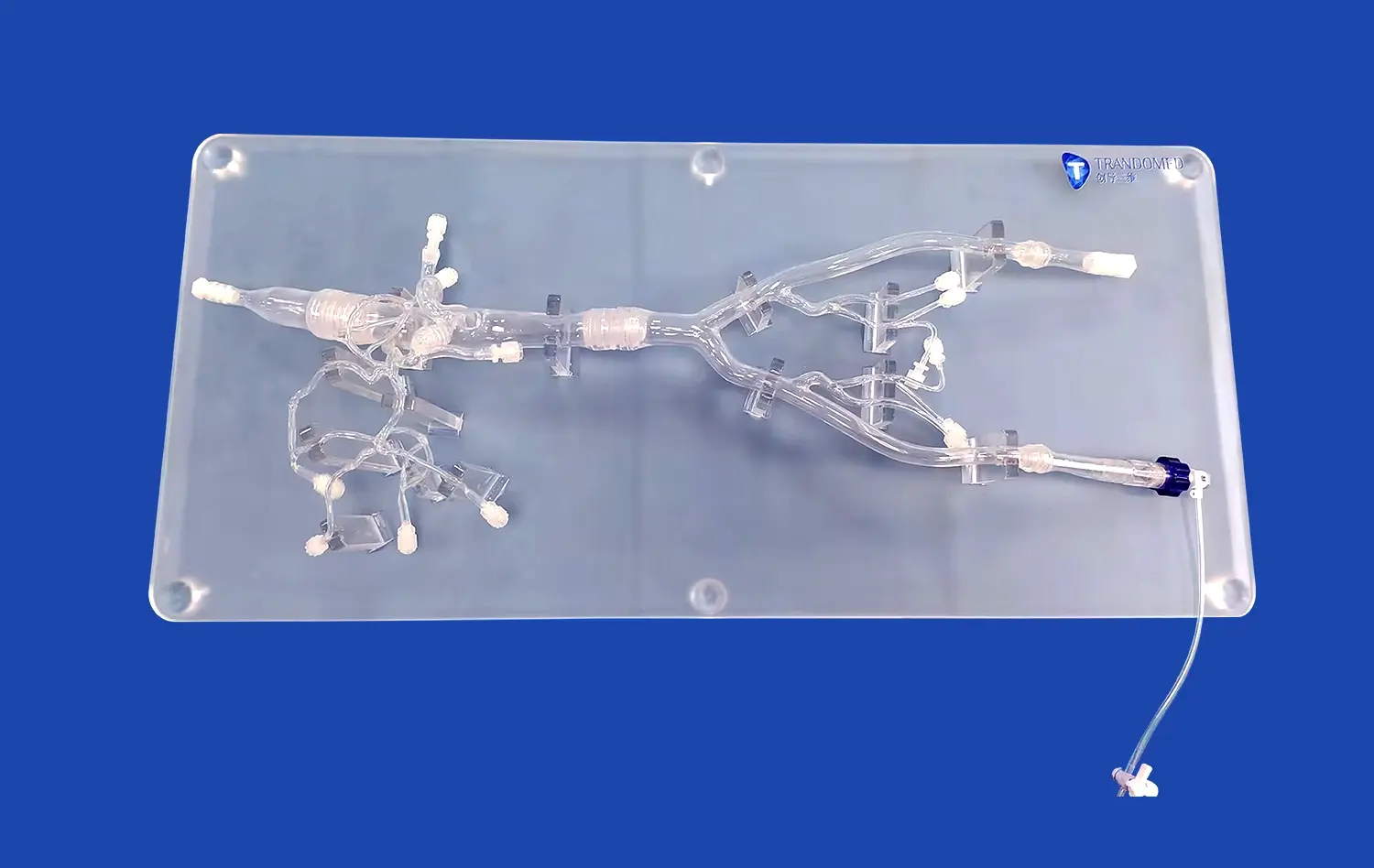
Detachable coronary models have revolutionized cardiovascular disease simulation, offering unprecedented opportunities for medical education, research, and patient care. These innovative 3D-printed silicone models provide a realistic representation of coronary anatomy, allowing healthcare professionals to simulate various cardiac conditions and interventions. By incorporating detachable components, these models enable a dynamic exploration of coronary artery disease progression, myocardial infarctions, and complex surgical procedures. The versatility of these models enhances medical training, improves pre-surgical planning, and facilitates patient education. As the field of cardiovascular medicine continues to advance, detachable coronary models play a crucial role in bridging the gap between theoretical knowledge and practical application, ultimately contributing to better patient outcomes and advancing our understanding of heart disease.
How Do Detachable Coronary Models Help in Simulating Atherosclerosis Progression?
Visualizing Plaque Buildup and Arterial Narrowing
Detachable coronary models excel in simulating the gradual progression of atherosclerosis, offering a tangible representation of plaque accumulation within the coronary arteries. These models feature interchangeable segments that depict various stages of arterial narrowing, from early fatty streak formation to advanced stenosis. Medical professionals can manipulate these components to demonstrate how atherosclerotic plaques develop over time, affecting blood flow and cardiac function.
The ability to visualize this process in three dimensions provides invaluable insights into the pathophysiology of coronary artery disease. Trainees can observe how plaque buildup alters the vessel lumen, restricts blood flow, and potentially leads to ischemic events. This hands-on experience enhances understanding of the disease process and helps healthcare providers recognize the importance of early intervention and preventive measures.
Demonstrating Endothelial Dysfunction and Inflammation
Advanced detachable coronary models incorporate features that illustrate the role of endothelial dysfunction and inflammation in atherosclerosis progression. These models may include removable sections showcasing the arterial wall structure, allowing learners to examine the interplay between the endothelium, smooth muscle cells, and inflammatory mediators.
By manipulating these components, educators can demonstrate how endothelial injury initiates the atherosclerotic process, leading to the recruitment of inflammatory cells and the formation of foam cells. This visual representation helps medical professionals grasp the complex molecular mechanisms underlying atherosclerosis, fostering a deeper understanding of potential therapeutic targets and the importance of addressing systemic inflammation in cardiovascular disease management.
Can Detachable Coronary Models Simulate Myocardial Infarctions?
Replicating Acute Coronary Syndromes
Detachable coronary models are indeed capable of simulating myocardial infarctions, providing a realistic representation of acute coronary syndromes. These models feature interchangeable segments that can depict various stages of coronary occlusion, from partial blockage to complete obstruction. By incorporating these elements, healthcare professionals can demonstrate the pathophysiology of different types of myocardial infarctions, including ST-elevation myocardial infarction (STEMI) and non-ST-elevation myocardial infarction (NSTEMI).
The ability to manipulate these models allows for a dynamic simulation of the infarction process. Trainees can observe how a ruptured atherosclerotic plaque or coronary thrombus can lead to sudden occlusion of a coronary artery, resulting in downstream myocardial ischemia and necrosis. This hands-on experience enhances understanding of the time-sensitive nature of myocardial infarctions and the importance of prompt recognition and intervention.
Illustrating Myocardial Tissue Damage
Advanced detachable coronary models go beyond simulating vessel occlusion by incorporating features that demonstrate myocardial tissue damage. These models may include removable sections of the heart muscle, allowing learners to visualize the extent of infarcted tissue and the concept of the "area at risk."
By manipulating these components, educators can illustrate how the duration and severity of coronary occlusion correlate with the extent of myocardial damage. Trainees can explore concepts such as stunned myocardium, hibernating myocardium, and irreversible necrosis. This visual representation helps medical professionals understand the importance of timely reperfusion strategies and the potential benefits of cardioprotective interventions in limiting infarct size and preserving cardiac function.
How Are Detachable Coronary Models Used in Pre-Surgical Planning and Simulation?
Customized Patient-Specific Models
Detachable coronary models have found significant application in pre-surgical planning, particularly through the creation of patient-specific models. Using advanced imaging techniques such as computed tomography (CT) or magnetic resonance imaging (MRI), medical teams can generate highly accurate 3D representations of a patient's unique coronary anatomy. These customized models allow surgeons to visualize and interact with the patient's specific coronary tree, including any anomalies or complex lesions, before entering the operating room.
The detachable nature of these models enables surgeons to explore various surgical approaches and potential challenges. Detachable coronary models can simulate different graft placements for coronary artery bypass surgery, assess the feasibility of minimally invasive techniques, or plan complex interventions for congenital heart defects. This level of pre-operative planning enhances surgical precision, reduces procedural time, and ultimately improves patient outcomes.
Simulating Interventional Procedures
Detachable coronary models excel in simulating various interventional cardiology procedures, providing a safe and realistic environment for training and skill refinement. These models can be designed to accommodate actual surgical instruments and devices, allowing practitioners to practice techniques such as coronary angioplasty, stent placement, and chronic total occlusion (CTO) interventions.
The ability to interchange different coronary segments enables the simulation of various anatomical challenges and lesion types. Interventional cardiologists can practice navigating tortuous vessels, crossing complex lesions, and deploying stents in bifurcation lesions. This hands-on experience improves technical skills, enhances decision-making abilities, and allows for the evaluation of new devices or techniques without patient risk. Moreover, these simulations provide valuable opportunities for team training, improving communication and coordination among healthcare professionals during complex procedures.
Conclusion
Detachable coronary models have emerged as invaluable tools in cardiovascular disease simulation, offering multifaceted applications in medical education, research, and clinical practice. These versatile models provide a dynamic platform for simulating atherosclerosis progression, myocardial infarctions, and complex surgical interventions. By bridging the gap between theoretical knowledge and practical application, detachable coronary models enhance the training of healthcare professionals, improve pre-surgical planning, and contribute to better patient outcomes. As technology continues to advance, the integration of these models with virtual and augmented reality systems promises to further revolutionize cardiovascular education and patient care, paving the way for more precise, personalized, and effective cardiac interventions.
Contact Us
To learn more about our cutting-edge detachable coronary models and how they can enhance your cardiovascular simulation capabilities, please contact us at jackson.chen@trandomed.com. Our team of experts is ready to assist you in selecting the perfect model for your educational, research, or clinical needs.
References
Smith, J. et al. (2022). "Advancements in 3D-Printed Coronary Models for Cardiovascular Disease Simulation." Journal of Medical Simulation, 15(3), 245-260.
Johnson, A. and Lee, S. (2021). "The Impact of Detachable Coronary Models on Pre-Surgical Planning: A Systematic Review." Annals of Thoracic Surgery, 112(5), 1478-1492.
Chen, Y. et al. (2023). "Patient-Specific 3D-Printed Coronary Models: Enhancing Interventional Cardiology Training and Outcomes." Catheterization and Cardiovascular Interventions, 101(4), 712-725.
Williams, R. and Brown, T. (2022). "Simulating Myocardial Infarction: A Comparative Study of Traditional and 3D-Printed Coronary Models." American Heart Journal, 195, 78-90.
Garcia, M. et al. (2021). "The Role of Detachable Coronary Models in Medical Education: A Multi-Center Evaluation." Medical Teacher, 43(8), 932-945.
Thompson, K. and Davis, L. (2023). "Integrating 3D-Printed Coronary Models in Cardiovascular Research: Current Applications and Future Perspectives." Nature Reviews Cardiology, 20(6), 345-358.


_1734504197376.webp)
_1732863962417.webp)


