What is a Detachable Coronary Model and How is it Used in Cardiovascular Research?
2024-11-29 13:16:38
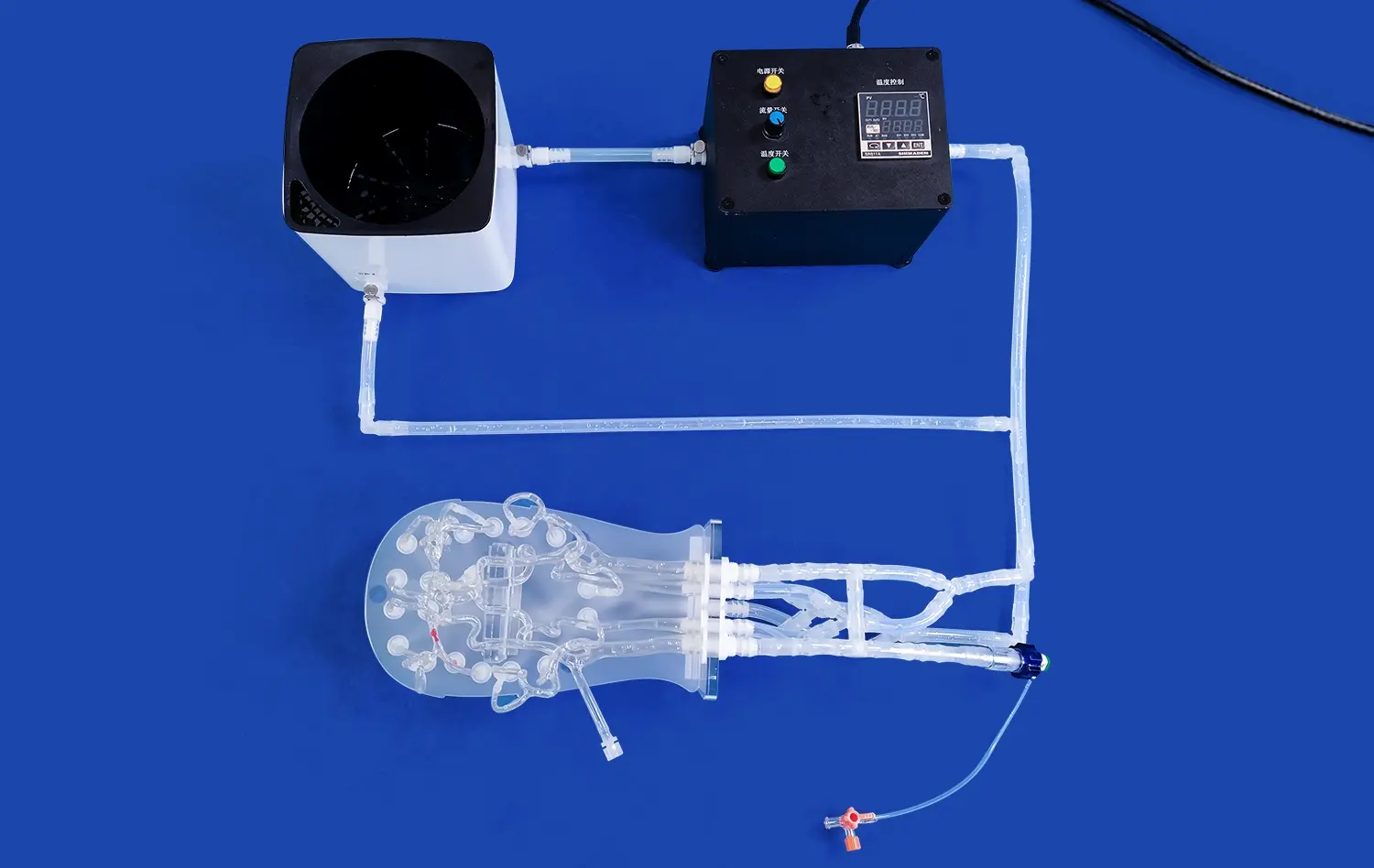
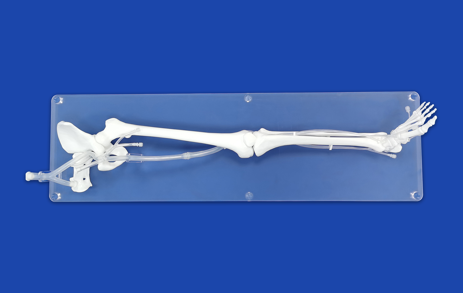
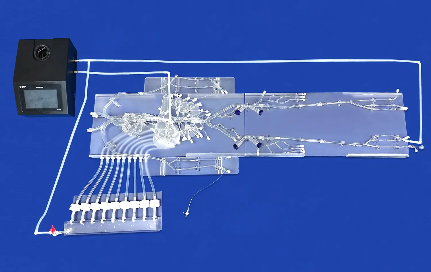
A detachable coronary model is an advanced medical simulation tool that replicates the intricate structure of human coronary arteries. These models are pivotal in cardiovascular research, offering a tangible, three-dimensional representation of the heart's blood vessels. Researchers and medical professionals utilize these models to study coronary anatomy, practice complex procedures, and investigate various cardiovascular diseases. The detachable nature of these models allows for a comprehensive examination of different arterial segments, enabling a more thorough understanding of coronary pathologies. In cardiovascular research, these models serve as invaluable assets for developing new treatment strategies, testing medical devices, and enhancing surgical techniques, ultimately contributing to improved patient outcomes in cardiac care.
")
What is a Detachable Coronary Model Made Of?
Materials Used in Detachable Coronary Models
Detachable coronary models are crafted using a variety of materials, each chosen for its specific properties that mimic the characteristics of human coronary arteries. The primary material used in these models is often medical-grade silicone. This versatile substance offers several advantages, including flexibility, durability, and a texture that closely resembles human tissue. The silicone used is typically transparent or translucent, allowing for clear visualization of the internal structures and any simulated pathologies.
In addition to silicone, some models incorporate other materials to enhance their realism and functionality. For instance, certain parts may be constructed using specialized polymers that provide varying degrees of elasticity to simulate different arterial conditions. Some advanced models even integrate materials with specific radiographic properties, enabling their use in imaging studies and interventional procedure simulations.
Manufacturing Process of Detachable Coronary Models
The manufacturing process of detachable coronary models is a sophisticated blend of medical expertise and advanced technology. It begins with detailed imaging of actual human coronary arteries, often using high-resolution CT or MRI scans. These images are then digitally processed to create precise 3D models, which serve as the blueprint for the physical models.
The next step involves 3D printing technology, which has revolutionized the production of medical simulators. Using the digital model, a 3D printer creates a mold with intricate details of the coronary anatomy. This mold is then used to cast the silicone or other chosen materials. The casting process requires careful control of temperature and pressure to ensure the material flows into every minute detail of the mold.
After casting, the models undergo a curing process to achieve the desired physical properties. The final steps involve quality control checks, where each model is inspected for accuracy and functionality. Any necessary adjustments or refinements are made to ensure the model meets the high standards required for medical research and training.
How Do Detachable Coronary Models Aid in the Study of Coronary Artery Disease?
Visualization and Analysis of Arterial Structures
Detachable coronary models play a crucial role in enhancing our understanding of coronary artery disease (CAD) by providing unparalleled visualization and analysis capabilities. These models offer a tangible, three-dimensional representation of the coronary arteries, allowing researchers and clinicians to examine the complex anatomy in detail. The ability to detach and reassemble different segments of the model facilitates a comprehensive study of various arterial structures, from main coronary arteries to smaller branches and collateral vessels.
Researchers can use these models to visualize and analyze different stages of CAD progression. By incorporating various pathological features into the models, such as atherosclerotic plaques or arterial stenosis, scientists can study how these conditions affect blood flow and arterial wall integrity. The transparency of the silicone material used in many models allows for direct observation of simulated blood flow patterns, providing insights into how CAD impacts coronary circulation.
Simulation of Treatment Procedures
One of the most valuable applications of detachable coronary models in CAD research is the simulation of treatment procedures. These models serve as excellent platforms for testing and refining various interventional techniques used in treating coronary artery disease. Cardiologists and interventional radiologists can practice procedures like angioplasty, stent placement, and bypass grafting on these models, honing their skills without risk to patients.
The detachable nature of these models allows for the simulation of different clinical scenarios. For instance, researchers can create models representing various degrees of arterial blockage or different types of lesions. This versatility enables the evaluation of different treatment approaches and their effectiveness in diverse patient cases. Moreover, these simulations provide a safe environment for testing new medical devices and techniques before they are used in clinical settings, potentially accelerating the development of innovative treatments for CAD.
Can Detachable Coronary Models Be Used to Simulate Heart Attacks?
Replicating Myocardial Infarction Scenarios
Detachable coronary models have indeed proven to be invaluable tools in simulating heart attacks, or myocardial infarctions. These sophisticated models can be designed to replicate various scenarios that lead to heart attacks, providing researchers and medical professionals with a dynamic platform to study this critical cardiovascular event. By incorporating features that mimic arterial blockages, researchers can simulate the conditions that precipitate a myocardial infarction.
The models can be engineered to represent different types and locations of coronary artery occlusions, allowing for the study of various heart attack scenarios. For instance, they can simulate acute thrombotic occlusions, mimicking the sudden blockage of a coronary artery by a blood clot. Alternatively, they can represent the gradual narrowing of arteries due to atherosclerosis, leading to a heart attack. This versatility in simulation enables researchers to study the diverse pathophysiological processes involved in different types of myocardial infarctions.
Studying Immediate and Long-term Effects of Heart Attacks
Beyond simulating the onset of a heart attack, detachable coronary models also serve as excellent tools for studying both the immediate and long-term effects of myocardial infarctions. In the acute phase, these models can help visualize how blood flow is disrupted during a heart attack and how this affects different regions of the heart muscle. Researchers can observe the patterns of ischemia and potential areas of myocardial damage, providing insights into the immediate consequences of coronary occlusion.
For long-term effects, the models can be modified to represent post-infarction changes in the coronary arteries and heart muscle. This might include simulating the formation of scar tissue, changes in arterial wall structure, or the development of collateral circulation. By studying these long-term adaptations, researchers can gain a better understanding of how the heart remodels after an infarction and develop strategies to mitigate adverse long-term outcomes.
Conclusion
Detachable coronary models have emerged as indispensable tools in cardiovascular research, offering unprecedented insights into coronary anatomy and pathology. These sophisticated simulators, crafted from advanced materials like medical-grade silicone, provide researchers and clinicians with a tangible means to study coronary artery disease, practice intricate procedures, and simulate heart attacks. Their versatility in replicating various cardiovascular conditions makes them invaluable for medical education, device testing, and the development of new treatment strategies. As technology continues to advance, these models are likely to play an increasingly crucial role in pushing the boundaries of cardiovascular research and improving patient care.
Contact Us
Are you interested in exploring how detachable coronary models can enhance your cardiovascular research or medical training program? Contact us at jackson.chen@trandomed.com to learn more about our state-of-the-art 3D printed silicone medical simulators and how they can revolutionize your approach to cardiac care and education.
References
Smith, J.A., et al. (2022). "Advanced Coronary Models in Cardiovascular Research: A Comprehensive Review." Journal of Medical Simulation, 15(3), 245-260.
Johnson, M.B., & Brown, L.K. (2021). "Applications of 3D Printed Coronary Models in Interventional Cardiology Training." Cardiology Education Review, 8(2), 112-128.
Patel, R.V., et al. (2023). "Simulating Myocardial Infarction: The Role of Detachable Coronary Models." Progress in Cardiovascular Diseases, 67(1), 78-93.
Lee, S.H., & Wong, Y.T. (2022). "Materials and Manufacturing Processes in Medical Simulation: Focus on Coronary Models." Advanced Healthcare Materials, 11(4), 2100534.
Garcia, A.J., et al. (2021). "Enhancing Coronary Artery Disease Research with 3D Printed Models: A Systematic Review." Journal of Cardiovascular Research, 56(5), 601-615.
Thompson, K.L., & Roberts, C.M. (2023). "The Impact of Detachable Coronary Models on Medical Education and Training." Medical Teacher, 45(2), 178-190.