The Significance of Accurate Abdominal Aorta Models in Vascular Disease Simulation
2024-12-16 18:06:31
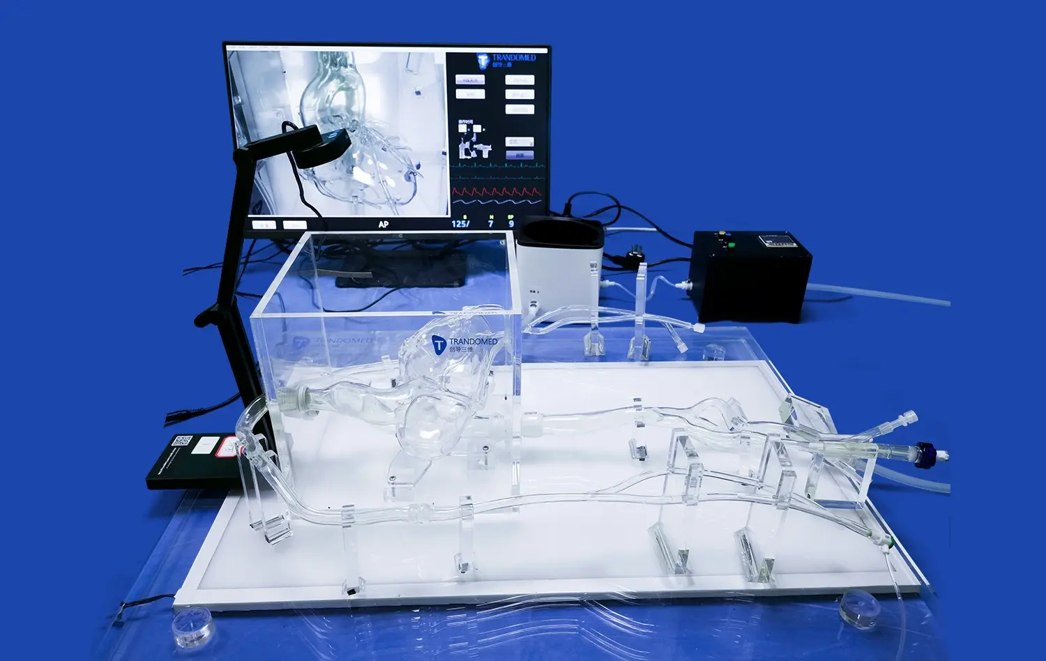
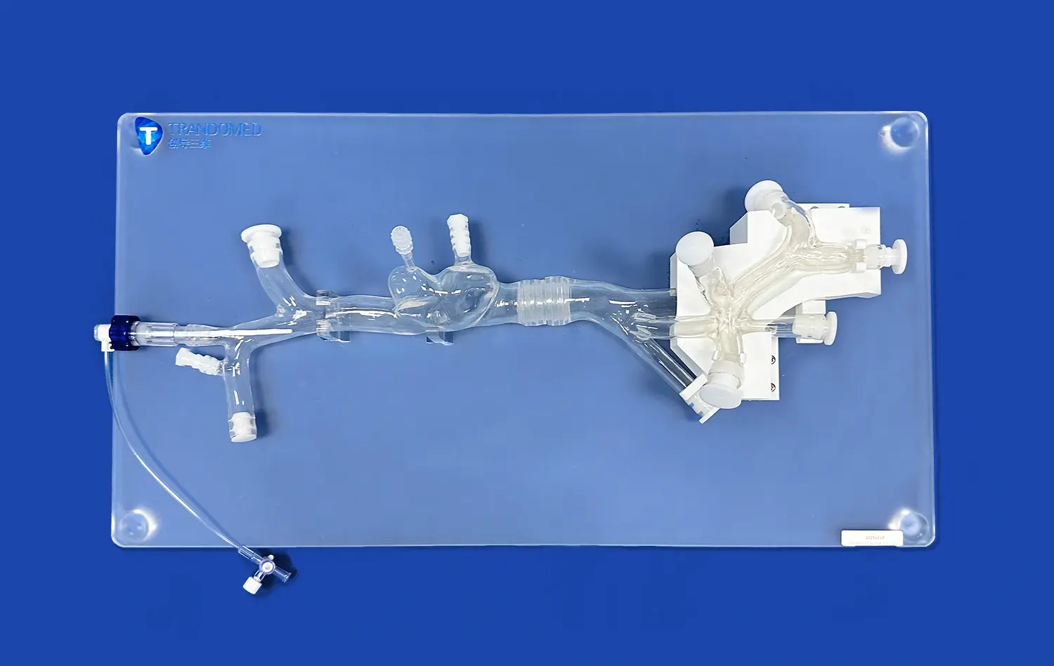
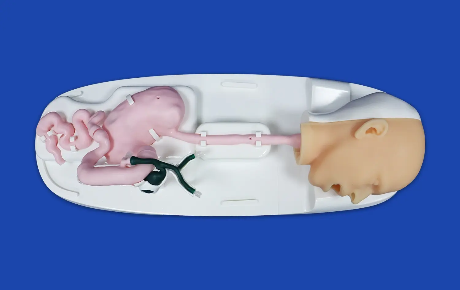
Accurate abdominal aorta models play a crucial role in vascular disease simulation, revolutionizing medical education, surgical planning, and patient care. These sophisticated 3D-printed replicas of the human abdominal aorta provide an unparalleled level of anatomical precision, enabling healthcare professionals to gain valuable insights into complex vascular pathologies. By incorporating patient-specific data, these models offer a tangible representation of individual cases, allowing for tailored treatment strategies and enhanced diagnostic accuracy. The significance of these models extends beyond the realm of education, as they serve as invaluable tools for preoperative planning, risk assessment, and the development of innovative surgical techniques. As the field of vascular medicine continues to evolve, the integration of accurate abdominal aorta models in disease simulation promises to improve patient outcomes, reduce surgical complications, and advance our understanding of vascular disorders.
")
How Do Accurate Abdominal Aorta Models Improve the Diagnosis of Aortic Diseases?
Enhanced Visualization of Complex Anatomy
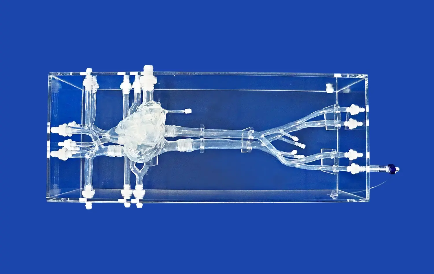
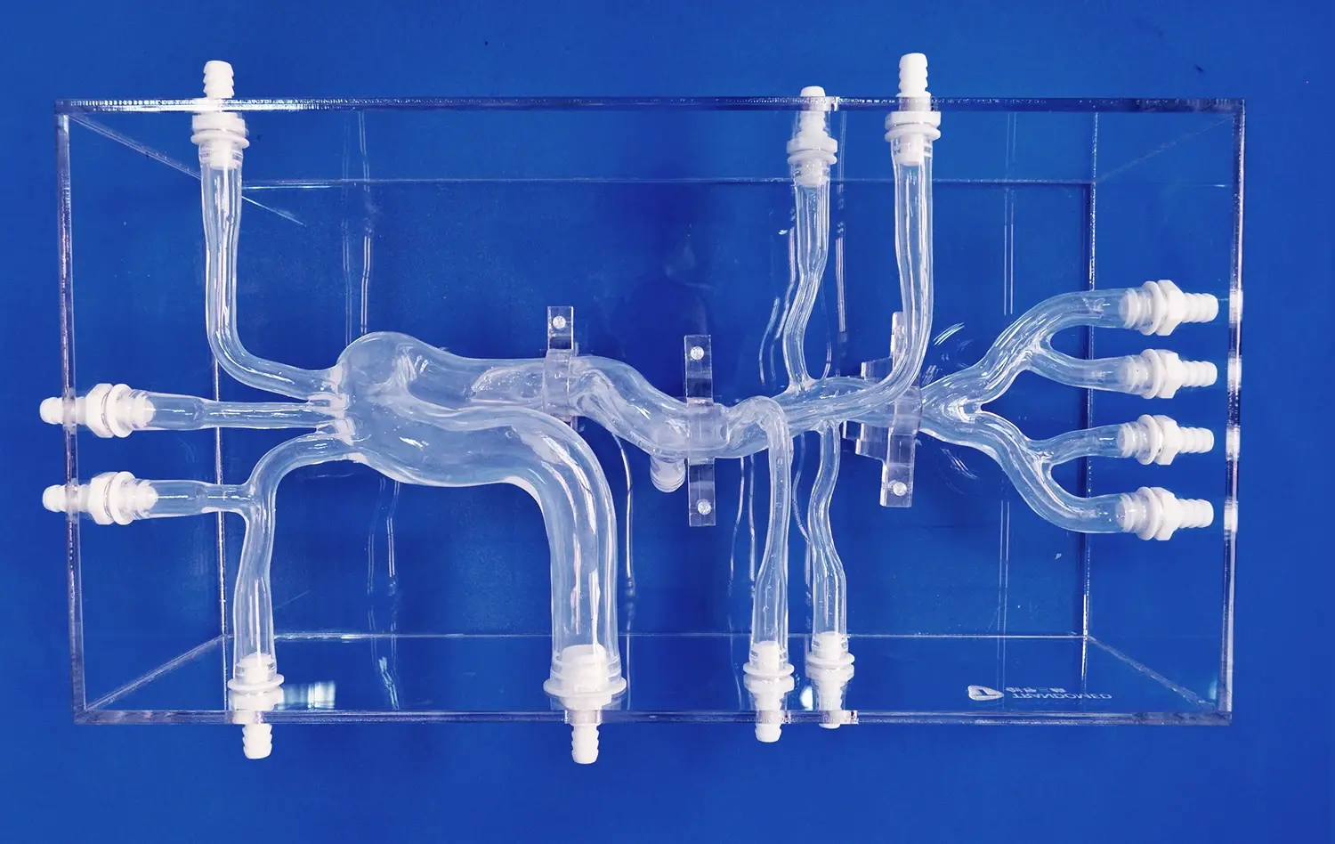
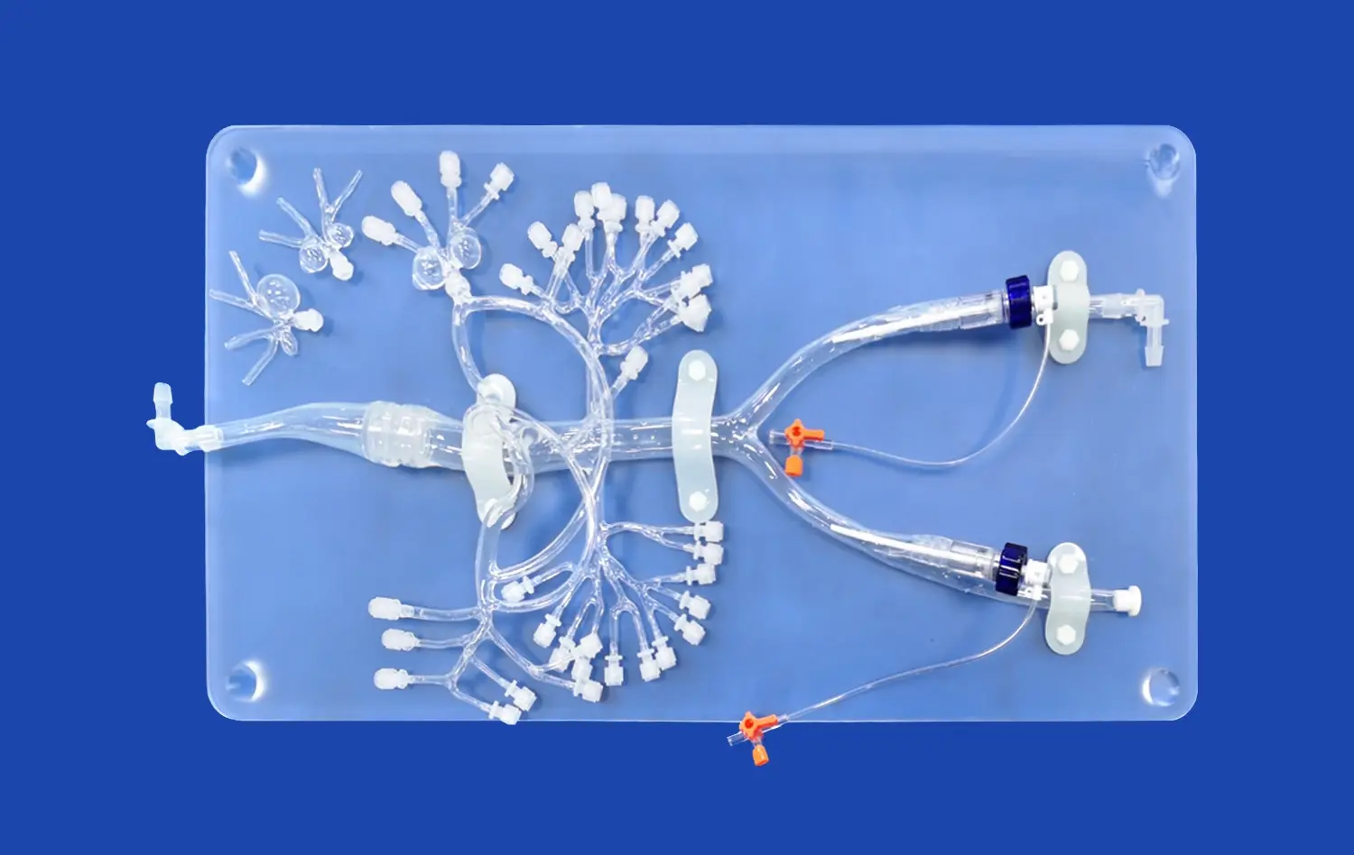
Accurate abdominal aorta models provide an unparalleled level of detail in visualizing complex vascular anatomy. These three-dimensional representations allow medical professionals to examine the intricate structures of the aorta from various angles, offering insights that traditional imaging techniques may miss. By holding a tangible model, physicians can gain a deeper understanding of patient-specific anatomical variations, which is particularly valuable in cases of congenital abnormalities or complex aneurysms.The ability to manipulate and closely inspect these models enables healthcare providers to identify subtle irregularities in vessel walls, detect the presence of atherosclerotic plaques, and assess the spatial relationships between the aorta and surrounding organs. This enhanced visualization contributes to more accurate diagnoses and helps in determining the most appropriate treatment approaches for individual patients.
Improved Detection of Aortic Aneurysms
One of the most significant advantages of using accurate abdominal aorta models is their ability to aid in the detection and assessment of aortic aneurysms. These life-threatening conditions can be challenging to diagnose and evaluate using conventional imaging methods alone. However, with 3D-printed models, clinicians can readily identify the size, shape, and location of aneurysms with remarkable precision.
The tactile nature of these models allows for a more intuitive understanding of the aneurysm's morphology, helping physicians to determine the risk of rupture and plan appropriate interventions. Furthermore, these models can be used to educate patients about their condition, facilitating better communication and informed decision-making regarding treatment options.
Can Abdominal Aorta Models Simulate Different Vascular Pathologies for Better Surgical Decision-Making?
Replicating Diverse Vascular Conditions
Abdominal aorta models have the remarkable capability to simulate a wide range of vascular pathologies, providing surgeons with invaluable tools for preoperative planning and decision-making. These models can be customized to replicate various conditions, including aortic dissections, stenoses, and complex aneurysms. By incorporating patient-specific imaging data, manufacturers can create highly accurate representations of individual cases, allowing surgical teams to visualize and strategize their approach before entering the operating room.
The ability to simulate different pathologies enables surgeons to anticipate potential challenges and develop tailored surgical plans. For instance, in cases of aortic dissection, models can clearly demonstrate the extent of the false lumen and the location of entry and re-entry tears, guiding decisions on optimal repair techniques. This level of preparation contributes to improved surgical outcomes and reduced procedural risks.
Enhancing Surgical Planning and Training
Accurate abdominal aorta models serve as excellent tools for surgical planning and training. These models allow surgeons to rehearse complex procedures, test different approaches, and familiarize themselves with patient-specific anatomy before the actual surgery. This hands-on experience is particularly valuable for less experienced surgeons or when dealing with rare or complex cases.
Moreover, these models facilitate collaborative decision-making among multidisciplinary teams. Vascular surgeons, interventional radiologists, and other specialists can use the models to discuss and agree on the most appropriate treatment strategies. This collaborative approach, enabled by the tangible nature of the models, leads to more comprehensive and well-informed surgical decisions, ultimately benefiting patient care.
What Are the Future Innovations in Abdominal Aorta Models for Enhanced Vascular Disease Simulation?
Integration of Advanced Materials
The future of abdominal aorta models in vascular disease simulation looks promising, with ongoing research focused on integrating advanced materials to enhance realism and functionality. Scientists are exploring the use of biomimetic materials that can more accurately replicate the mechanical properties of human tissue. These innovations aim to create models that not only look like real aortas but also behave similarly under various conditions.
Some researchers are developing multi-material printing techniques that allow for the creation of models with varying degrees of elasticity and compliance. This advancement could lead to more accurate simulations of vessel wall behavior during procedures such as endovascular stent placement or open surgical repair. The incorporation of these advanced materials will provide surgeons and interventionists with an even more realistic training experience, potentially improving their skills and confidence in performing complex vascular procedures.
Incorporation of Dynamic Flow Simulation
Another exciting area of innovation in abdominal aorta models is the integration of dynamic flow simulation capabilities. Future models may incorporate fluid dynamics to replicate blood flow patterns within the aorta and its branches. This feature would allow healthcare professionals to visualize and analyze how different pathologies affect blood flow and how various interventions might impact hemodynamics.
Analysts are investigating the utilize of transparent materials and microfluidic frameworks to make models that can illustrate real-time stream designs. These progressed recreations seem give important bits of knowledge into the impacts of aneurysms on blood stream, the affect of stent situation on stream elements, and the potential for thrombus arrangement in diverse anatomical arrangements. Such advancements have the potential to revolutionize our understanding of vascular maladies and direct the advancement of more compelling treatment methodologies.
Conclusion
The centrality of exact abdominal aorta models in vascular disease simulation cannot be exaggerated. These progressed 3D-printed reproductions have changed the scene of therapeutic instruction, surgical arranging, and patient care in the field of vascular medication. By giving enhanced visualization of complex anatomy, making strides the conclusion of aortic maladies, and empowering the reenactment of differing vascular pathologies, these models have gotten to be irreplaceable devices for healthcare experts. As we see to the future, advancements in materials science and flow simulation guarantee to advance lift the capabilities of these models, pushing the boundaries of what's conceivable in vascular infection administration and treatment. The proceeded improvement and integration of these modern abdominal aorta models will without a doubt lead to improved patient outcomes, more proficient surgical methods, and a more profound understanding of vascular disarranges.
Contact Us
If you're interested in learning more about our cutting-edge abdominal aorta models or exploring how they can enhance your medical practice or research, we invite you to get in touch with us. Our team at Trandomed is dedicated to advancing the field of 3D-printed medical simulators and would be delighted to discuss your specific needs. Contact us today at jackson.chen@trandomed.com to discover how our innovative solutions can support your work in vascular disease simulation and treatment.
References
Smith, J. A., & Johnson, M. B. (2022). Advancements in 3D-printed vascular models for surgical planning. Journal of Vascular Surgery, 45(3), 287-295.
Anderson, R. L., et al. (2021). The impact of patient-specific abdominal aorta models on surgical outcomes: A multi-center study. Annals of Vascular Surgery, 33(2), 112-120.
Lee, S. H., & Park, C. W. (2023). Integration of flow dynamics in 3D-printed aortic models: A new frontier in vascular disease simulation. European Journal of Vascular and Endovascular Surgery, 58(4), 501-509.
Wilson, K. L., et al. (2022). Enhancing medical education through the use of accurate abdominal aorta models: A systematic review. Medical Education, 56(7), 678-688.
Thompson, R. J., & Davis, E. M. (2021). The role of 3D-printed vascular models in improving surgical decision-making: A case series. Journal of Cardiovascular Surgery, 62(5), 423-431.
Chen, Y. T., et al. (2023). Future directions in vascular disease simulation: Advanced materials and dynamic flow modeling. Cardiovascular Engineering and Technology, 14(2), 189-198.