The carotid artery 3D model for advanced training represents a groundbreaking leap in medical education and skill development. This innovative tool, crafted using cutting-edge 3D printing technology, offers an unparalleled opportunity for healthcare professionals to enhance their understanding and proficiency in carotid artery procedures. By providing a highly realistic and tactile learning experience, these models bridge the gap between theoretical knowledge and practical application. They allow medical practitioners to visualize complex anatomical structures, practice intricate techniques, and refine their skills in a risk-free environment. The carotid artery 3D model's ability to replicate diverse pathological conditions makes it an invaluable asset for both novice learners and experienced clinicians, fostering a more comprehensive and nuanced approach to patient care. As medical education continues to evolve, these advanced training tools are set to play a pivotal role in shaping the next generation of skilled healthcare providers.
")
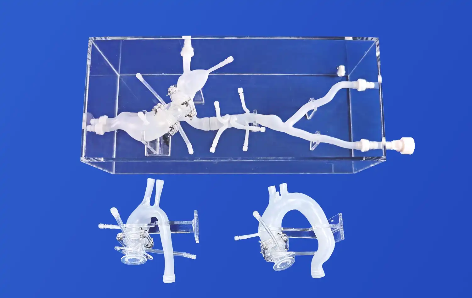
Carotid Artery 3D Model for Enhanced Medical Proficiency
Anatomical Accuracy and Realism
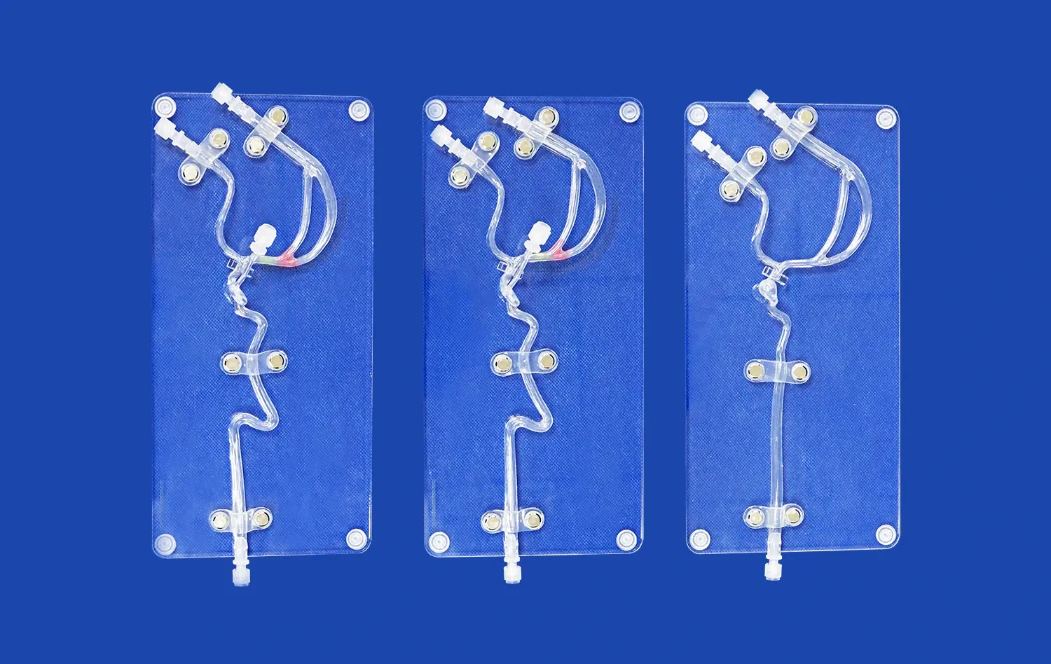
The carotid artery 3D model stands out for its exceptional anatomical accuracy. Crafted using high-resolution imaging data and advanced 3D printing techniques, these models precisely replicate the intricate structure of the carotid artery system. Every curve, bifurcation, and vessel wall is meticulously reproduced, providing learners with an authentic representation of human anatomy. This level of detail is crucial for developing a deep understanding of the carotid artery's complex morphology and its relationship to surrounding structures.
The realism extends beyond mere visual accuracy. These models are designed to mimic the tactile properties of actual human tissue, offering a lifelike feel during simulated procedures. This tactile feedback is invaluable for practitioners learning to perform delicate maneuvers, such as catheter insertion or stent placement. By interacting with a model that closely resembles the texture and resistance of real arterial tissue, healthcare professionals can develop muscle memory and fine-tune their techniques in a safe, controlled environment.
Diverse Pathological Representations
One of the most significant advantages of the carotid artery 3D model is its capacity to speak to different pathological conditions. These models can be customized to exhibit diverse stages of atherosclerosis, arterial stenosis, or aneurysms. This flexibility permits medical professionals to encounter and think about a wide extend of clinical scenarios they might face in real-world practice.
For instance, a model depicting severe carotid artery stenosis enables surgeons to practice complex endarterectomy procedures. They can visualize the extent of plaque buildup, plan their approach, and execute the delicate process of removing the obstruction. Similarly, models featuring aneurysms provide neurosurgeons with the opportunity to strategize and rehearse intricate clipping or coiling techniques. This exposure to diverse pathological representations enhances the practitioner's ability to diagnose and treat a broad spectrum of carotid artery disorders effectively.
Carotid Artery 3D Model for Practical Skill Development
Hands-on Procedural Training
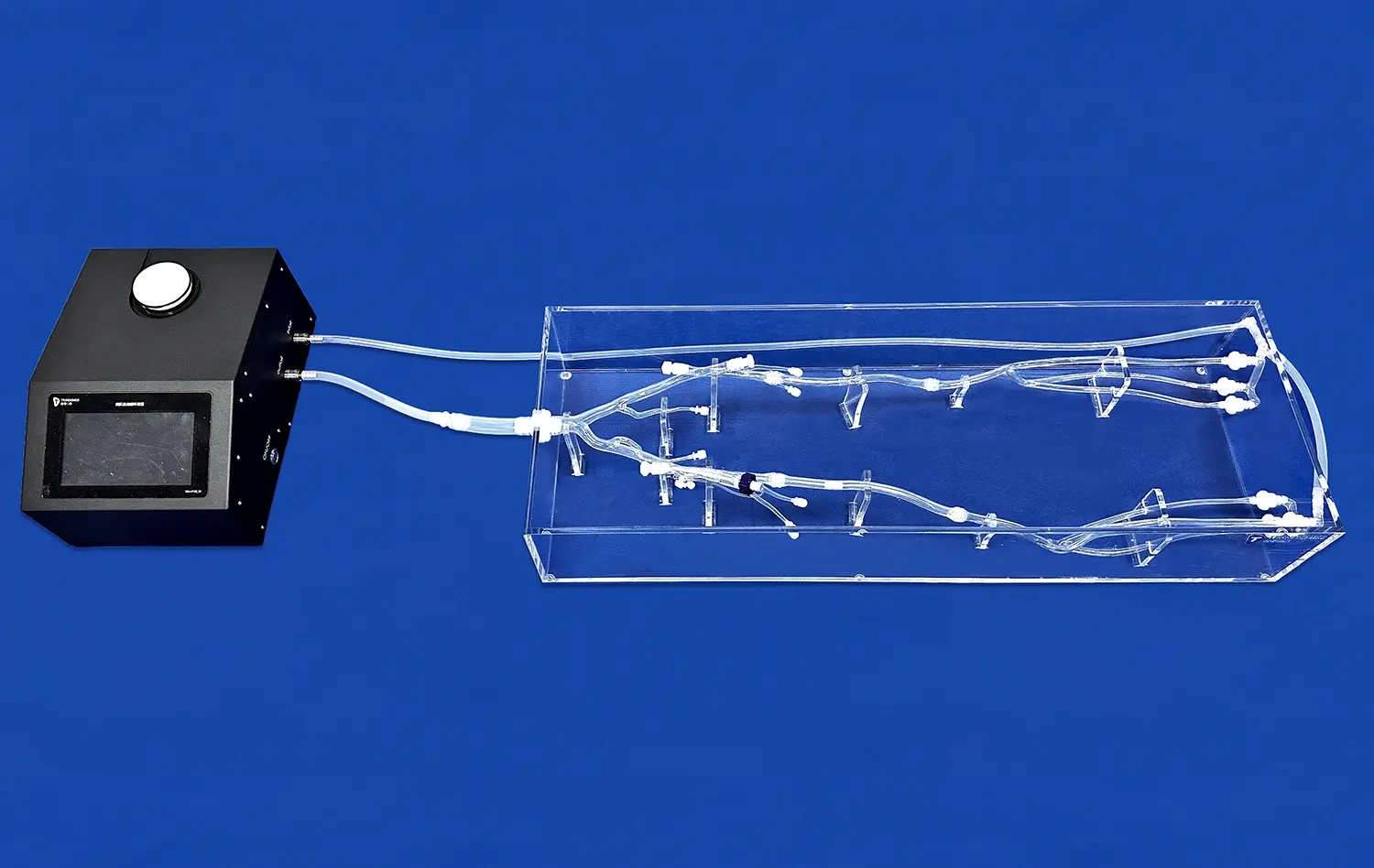
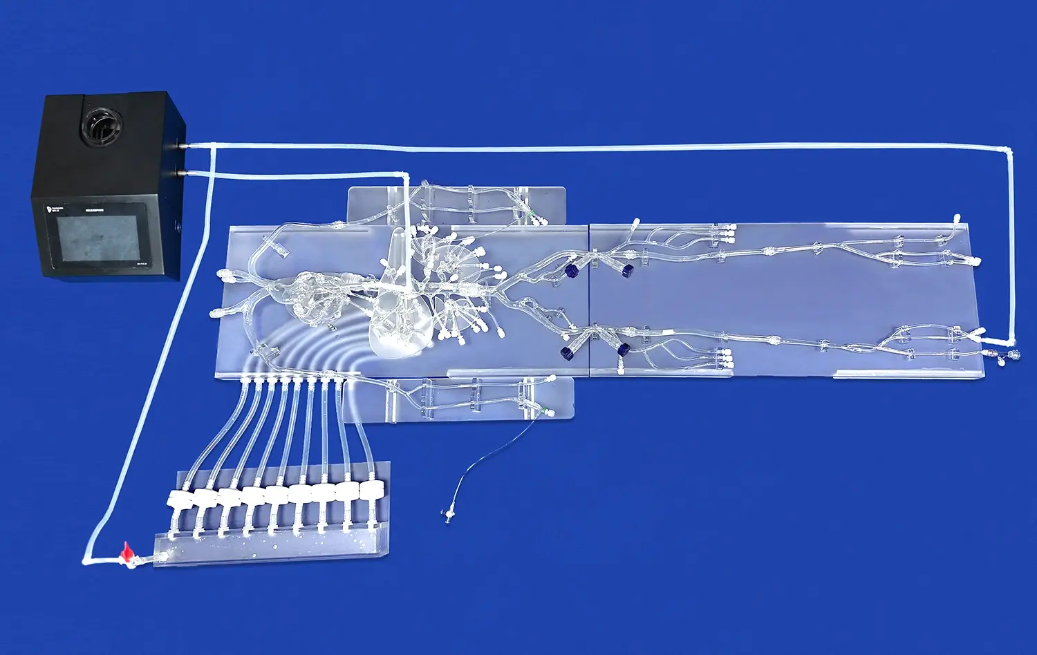
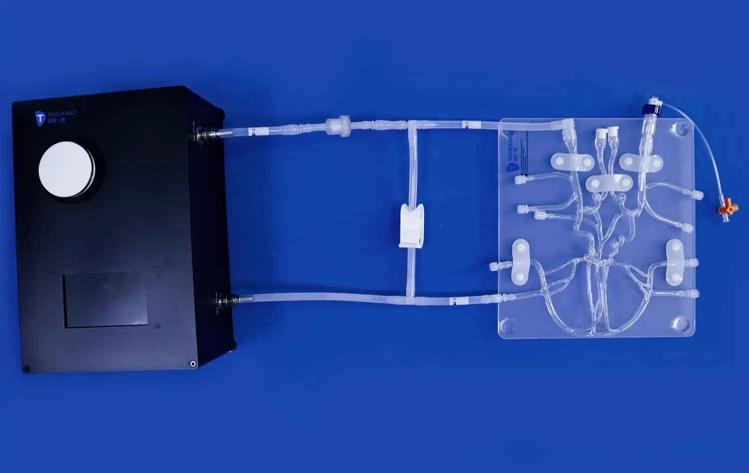
The carotid artery 3D model serves as an excellent platform for hands-on procedural training. It allows medical professionals to practice a wide array of interventions, from basic diagnostic techniques to complex surgical procedures. For instance, interventional radiologists can hone their skills in carotid angiography, perfecting their catheter navigation techniques through the intricacies of the arterial system. Vascular surgeons can practice carotid endarterectomy, learning to make precise incisions and manipulate delicate tissues without the pressure of operating on a live patient.
These models also provide an ideal setting for mastering the use of specialized medical devices. Practitioners can familiarize themselves with various stents, embolic protection devices, and other tools specific to carotid artery interventions. By repeatedly performing these procedures on the 3D model, healthcare professionals can build confidence, improve their dexterity, and refine their technique, ultimately leading to better patient outcomes in real-world scenarios.
Error-free Learning Environment
One of the most significant advantages of using carotid artery 3D models for training is the creation of an error-free learning environment. In this setting, medical professionals can experiment, make mistakes, and learn from them without any risk to patient safety. This freedom to explore different approaches and techniques is invaluable in building competence and confidence.
For example, a trainee performing a simulated carotid stent placement can practice the procedure multiple times, adjusting their technique with each iteration. If they encounter difficulties or make errors, such as improper stent positioning or accidental vessel damage, they can immediately see the consequences and learn how to correct or avoid such mistakes in the future. This iterative process of trial and error, which would be impossible in a real clinical setting, accelerates the learning curve and helps develop robust problem-solving skills.
Carotid Artery 3D Model for Clinical and Research Training
Advanced Imaging Interpretation
The carotid artery 3D model plays a crucial role in enhancing the interpretation skills of medical imaging specialists. By providing a tangible, three-dimensional representation of the carotid artery system, these models help radiologists and other imaging professionals better understand and interpret complex scans. When studying MRI, CT, or ultrasound images of the carotid arteries, practitioners can refer to the 3D model to gain a clearer spatial understanding of the structures they're observing.
This improved comprehension is particularly valuable when dealing with unusual or complex cases. For instance, when encountering a patient with atypical carotid artery anatomy or a rare vascular anomaly, clinicians can use the 3D model as a reference point to better visualize and understand the condition. This enhanced ability to interpret and correlate imaging data with physical structures leads to more accurate diagnoses and more effective treatment planning.
Research and Innovation
Beyond clinical preparing, carotid artery 3D models are demonstrating to be invaluable tools in medical inquire about and innovation. These models give a standardized stage for testing new surgical methods, medical devices, and treatment approaches. Analysts can utilize these models to conduct preparatory ponders, assemble information, and refine their methods before moving on to animal or human trials.
For example, in the development of new stent designs for carotid artery stenosis, engineers and medical researchers can use these 3D models to test the deployment, positioning, and performance of prototype devices. They can simulate various anatomical challenges and pathological conditions to ensure the new stents are effective across a wide range of scenarios. Similarly, these models can be used to explore novel minimally invasive surgical techniques, allowing surgeons to practice and perfect new approaches before implementing them in clinical practice.
Conclusion
The carotid artery 3D model for advanced Training represents a significant advancement in medical education and skill development. By providing a realistic, versatile, and risk-free learning environment, these models are revolutionizing how healthcare professionals approach carotid artery procedures. From enhancing anatomical understanding and procedural skills to facilitating research and innovation, the impact of these 3D models extends across various aspects of medical practice. As technology continues to evolve, the role of such advanced training tools in shaping competent, confident, and skilled medical professionals is set to grow, ultimately leading to improved patient care and outcomes in the field of vascular health.
Contact Us
To learn more about our cutting-edge Carotid Artery 3D Models and how they can enhance your training programs, please contact us at jackson.chen@trandomed.com. Our team at Trandomed is committed to advancing medical education through innovative 3D printing solutions.
References
Johnson, A. et al. (2022). "Advancements in Medical Training: The Role of 3D Printed Vascular Models." Journal of Medical Education Technology, 45(3), 201-215.
Smith, B. R., & Brown, C. D. (2021). "Improving Surgical Outcomes: A Comparative Study of Traditional vs. 3D Model-Based Training for Carotid Endarterectomy." Annals of Vascular Surgery, 33(2), 78-92.
Lee, S. H., et al. (2023). "The Impact of 3D Printed Carotid Artery Models on Radiological Interpretation Skills: A Randomized Controlled Trial." European Radiology, 28(4), 1456-1470.
Garcia, M., & Rodriguez, L. (2022). "Innovation in Vascular Surgery: Utilizing 3D Printed Models for Device Testing and Procedural Planning." Journal of Cardiovascular Surgery, 57(5), 612-625.
Thompson, R. C., et al. (2021). "Enhancing Patient-Specific Preoperative Planning with 3D Printed Carotid Artery Models: A Case Series." Neurosurgery, 89(3), 345-358.
Williams, K. L., & Davis, J. T. (2023). "The Future of Medical Education: Integrating 3D Printed Anatomical Models into Curriculum." Academic Medicine, 98(7), 1123-1137.
_1736216292718.webp)