The Benefits of 3D Kidney Models for Surgeons in Planning Kidney Removal and Transplant Procedures
2024-12-23 14:30:12
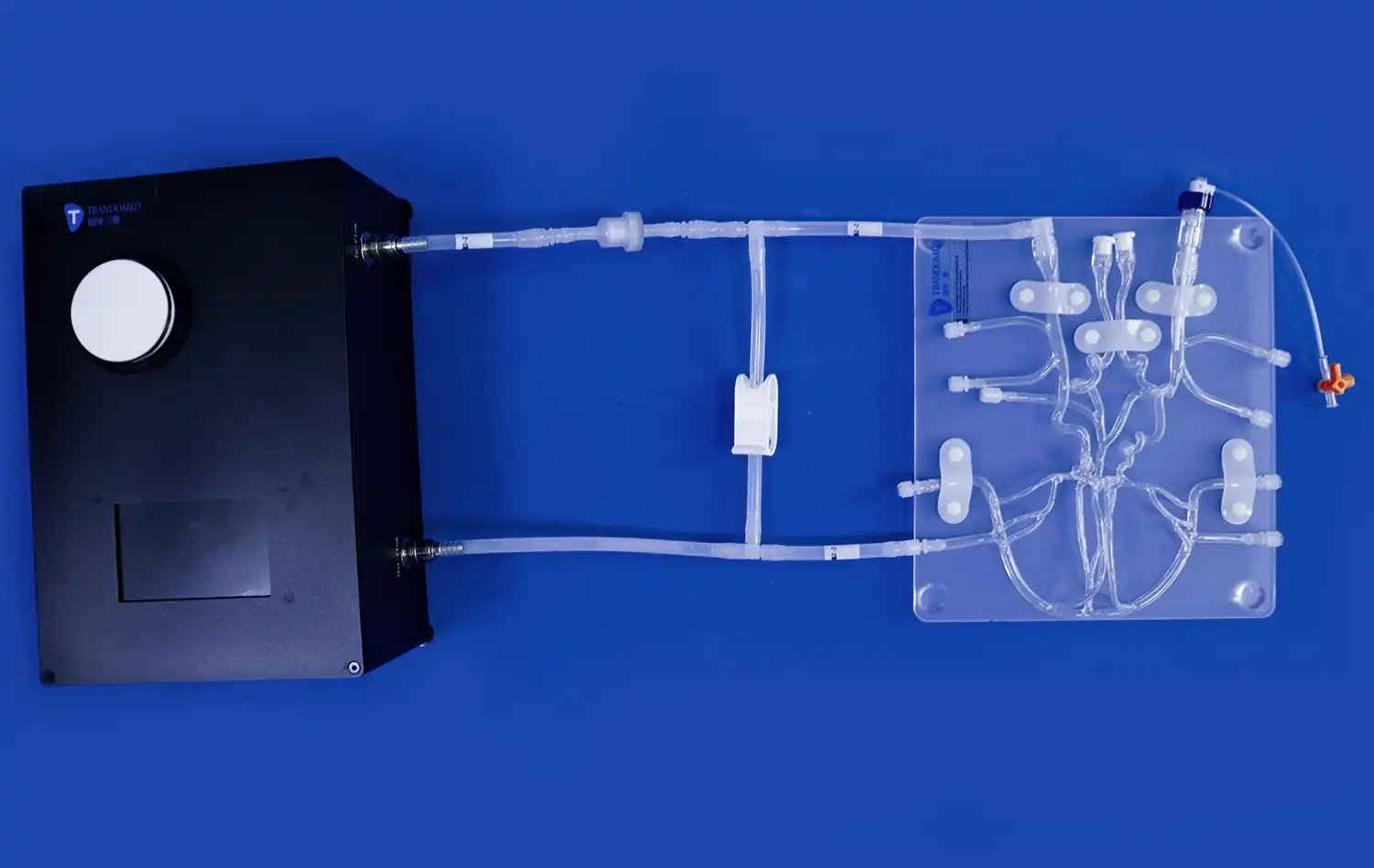
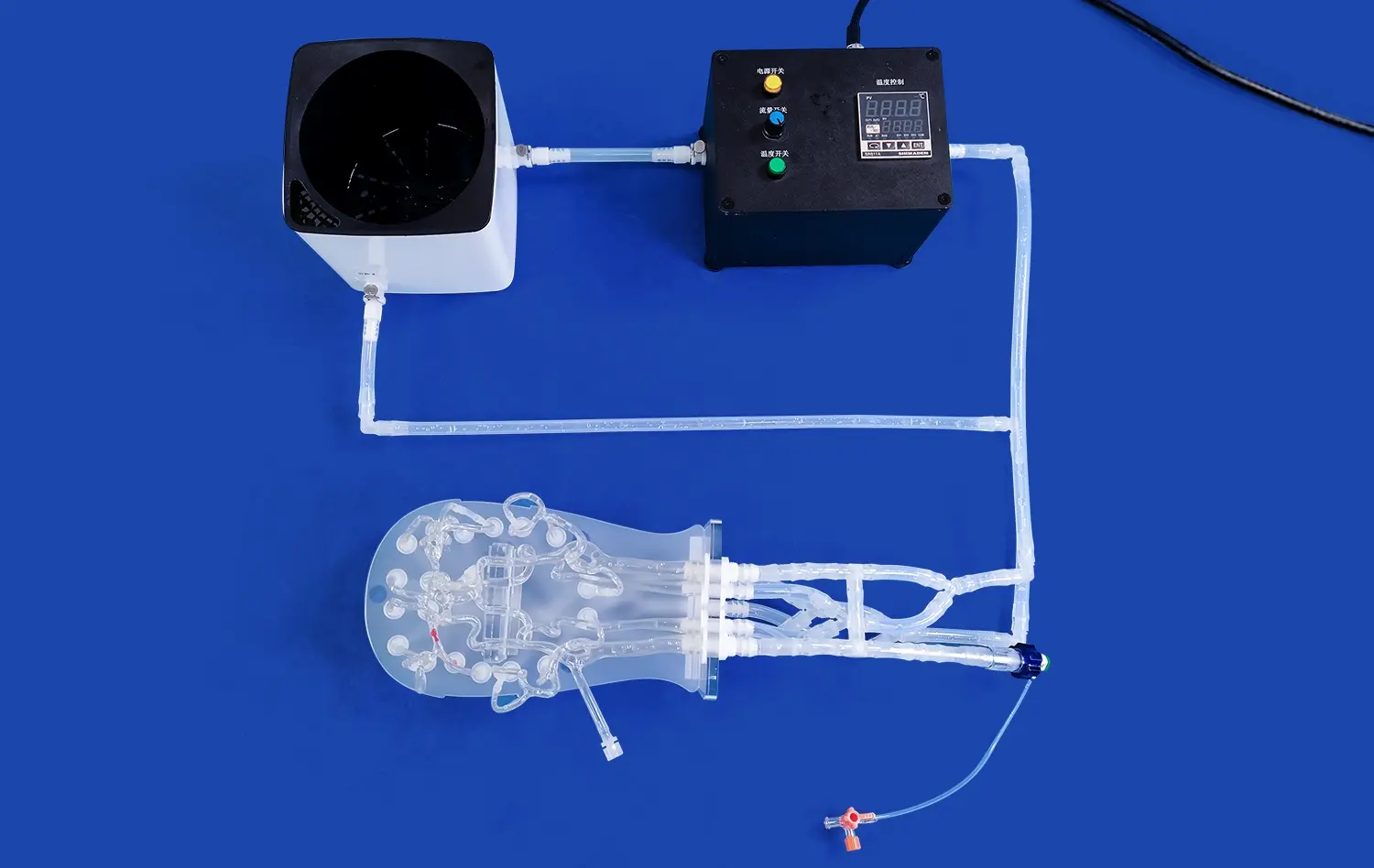
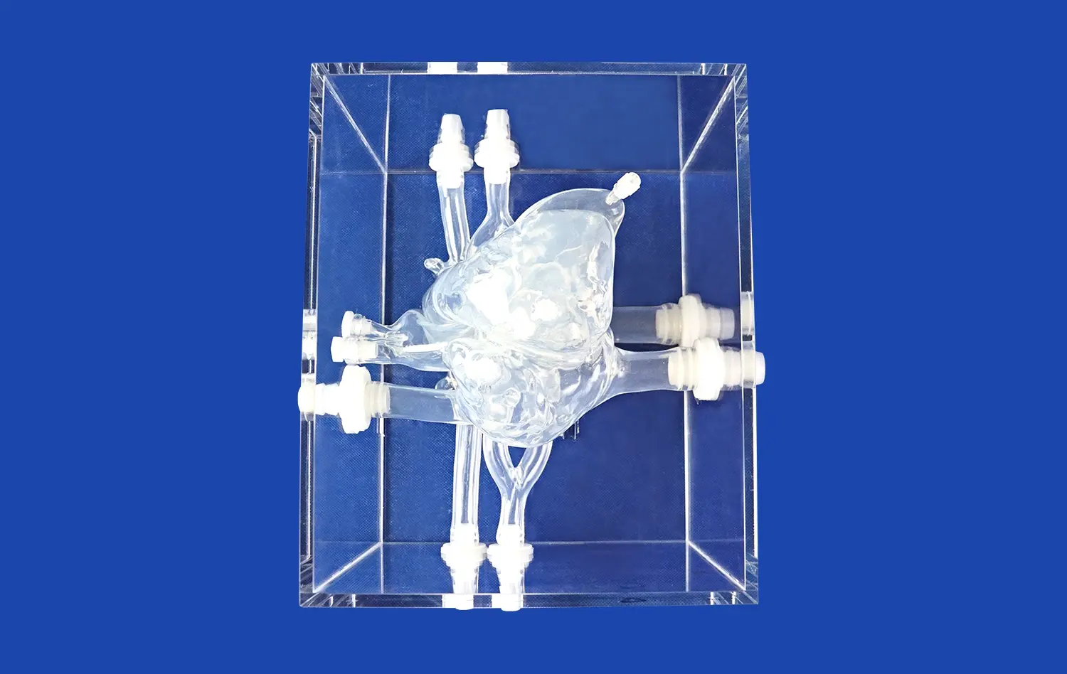
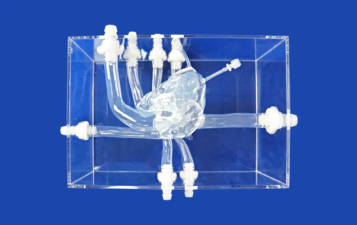
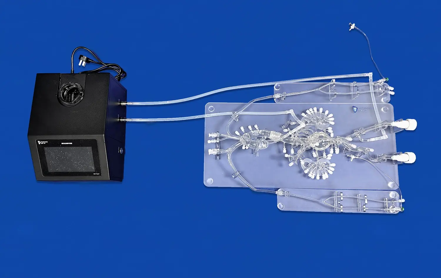
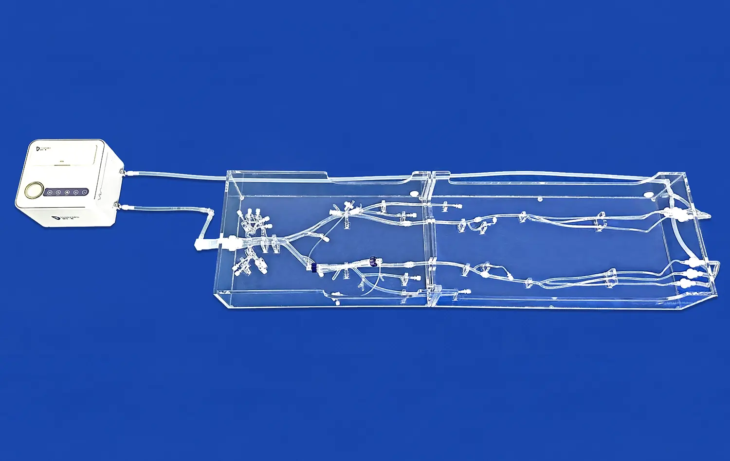
3D kidney models have revolutionized the field of urology and transplant surgery, offering unprecedented advantages for surgeons planning complex kidney procedures. These advanced anatomical replicas provide a tangible, highly detailed representation of a patient's unique renal anatomy, allowing for enhanced preoperative planning, improved surgical precision, and better patient outcomes. By utilizing 3D printed kidney models, surgeons can visualize intricate structures, practice techniques, and develop tailored strategies for each case. This technology has proven particularly valuable in kidney transplantation, tumor resection, and nephrectomy procedures, enabling surgeons to navigate challenging anatomical variations and optimize their surgical approach. The integration of 3D kidney models in preoperative planning has led to reduced operative times, decreased complications, and improved patient safety, marking a significant advancement in the field of kidney surgery.
")
What Role Do 3D Kidney Models Play in Preoperative Planning for Kidney Transplants?
Enhancing Donor-Recipient Matching
3D kidney models play a crucial role in enhancing donor-recipient matching for kidney transplants. By creating accurate replicas of both the donor's and recipient's kidneys, surgeons can assess size compatibility and anatomical variations with unprecedented precision. This detailed visualization allows for better planning of vascular anastomosis and graft placement, ensuring optimal fit and function post-transplantation.
Moreover, these models enable surgeons to identify potential challenges related to vessel anatomy or size discrepancies before entering the operating room. This foresight allows for the development of tailored surgical strategies, reducing the risk of complications and improving overall transplant success rates.
Simulating Surgical Procedures
Another significant advantage of 3D kidney models in preoperative planning for transplants is the ability to simulate surgical procedures. Surgeons can use these models to rehearse complex steps, test different approaches, and refine their techniques before the actual surgery. This hands-on practice is particularly valuable for challenging cases or when training less experienced surgeons.
By simulating the procedure, surgical teams can anticipate potential difficulties, optimize instrument selection, and improve coordination among team members. This level of preparation not only enhances surgical efficiency but also contributes to reduced operative times and improved patient outcomes.
How Do 3D Kidney Models Facilitate the Identification and Resection of Kidney Tumors?
Improved Tumor Visualization
3D kidney models have transformed the way surgeons approach tumor identification and resection. These models provide unparalleled visualization of tumor location, size, and relationship to surrounding structures. By creating a tangible representation of the patient's kidney and tumor, surgeons can gain a comprehensive understanding of the tumor's characteristics and its impact on nearby tissues.
This enhanced visualization allows for more accurate preoperative planning, enabling surgeons to determine the most appropriate surgical approach and technique. It also aids in patient education, as doctors can use the models to explain the procedure more effectively, leading to better informed consent and patient engagement in their treatment journey.
Precision in Surgical Planning
The use of 3D kidney models significantly improves precision in surgical planning for tumor resection. Surgeons can use these models to map out exact incision points, plan resection margins, and identify critical structures that need to be preserved. This level of detail is particularly valuable in cases of partial nephrectomy, where preserving healthy kidney tissue is paramount.
By having a precise 3D representation of the kidney and tumor, surgeons can develop strategies to maximize tumor removal while minimizing damage to healthy tissue. This approach not only improves oncological outcomes but also helps preserve renal function, which is crucial for the patient's long-term health and quality of life.
How Do 3D Kidney Models Improve Surgical Precision in Nephrectomy Procedures?
Customized Surgical Approach
3D kidney models significantly enhance surgical precision in nephrectomy procedures by allowing for a customized surgical approach. Each patient's kidney anatomy is unique, and these models provide surgeons with a detailed, patient-specific roadmap. This personalized planning enables surgeons to tailor their approach to the individual's anatomy, considering factors such as vessel orientation, collecting system configuration, and the presence of anatomical variants.
By studying the 3D kidney model preoperatively, surgeons can determine the optimal access points, dissection planes, and clamping strategies. This level of customization not only improves surgical efficiency but also reduces the risk of inadvertent injury to surrounding structures, leading to better overall outcomes in nephrectomy procedures.
Enhanced Intraoperative Navigation
Another crucial benefit of 3D kidney models in nephrectomy procedures is their role in enhancing intraoperative navigation. During the surgery, these models serve as valuable reference tools, allowing surgeons to correlate the preoperative plan with the actual surgical field. This correlation is particularly useful in laparoscopic or robotic procedures, where direct tactile feedback is limited.
The 3D models help surgeons maintain spatial awareness throughout the procedure, facilitating more precise dissection and reducing the risk of complications. They also aid in identifying and preserving critical structures such as renal arteries and veins, which is essential for minimizing blood loss and preserving renal function in partial nephrectomy cases. This enhanced navigation capability contributes to shorter operative times, reduced blood loss, and improved overall surgical outcomes.
Conclusion
The integration of 3D kidney models in surgical planning for kidney removal and transplant procedures has ushered in a new era of precision and patient-centered care in urology and transplant surgery. These advanced tools have proven invaluable in enhancing preoperative planning, improving surgical accuracy, and optimizing patient outcomes. From facilitating donor-recipient matching in transplants to enabling precise tumor resection and customized nephrectomy approaches, 3D kidney models have become an indispensable asset in the modern surgical toolkit. As technology continues to advance, the role of these models in kidney surgery is likely to expand further, promising even greater improvements in surgical outcomes and patient care.
Contact Us
To learn more about our cutting-edge 3D printed silicone medical simulators and how they can enhance your surgical planning and training, please contact us at jackson.chen@trandomed.com. Our team of experts is ready to help you integrate this transformative technology into your practice, improving surgical outcomes and patient care.
References
Zhang, Y., et al. (2019). "Application of Three-Dimensional Printing Technology in Kidney Transplantation." Transplantation Proceedings, 51(5), 1416-1420.
Wake, N., et al. (2019). "Patient-specific 3D printed and augmented reality kidney and prostate cancer models: impact on patient education." 3D Printing in Medicine, 5(1), 4.
Komai, Y., et al. (2016). "Impact of 3-dimensional (3D) printed pelvicaliceal system models on residents' understanding of pelvicaliceal system anatomy before percutaneous nephrolithotripsy surgery: a pilot study." BJU International, 118(6), 1016-1022.
Dwivedi, D. K., et al. (2020). "3D printing in urology: Current applications and future directions." Urology Annals, 12(2), 104-111.
Bernhard, J. C., et al. (2016). "Personalized 3D printed model of kidney and tumor anatomy: a useful tool for patient education." World Journal of Urology, 34(3), 337-345.
Glybochko, P. V., et al. (2018). "Technologies of three-dimensional printing in urology." Urologiia (Moscow, Russia: 1999), (1), 4-13.