Labeled Cranial Nerves Models: A Comprehensive Tool for Neurology and Anatomy Education
2025-01-07 09:08:37
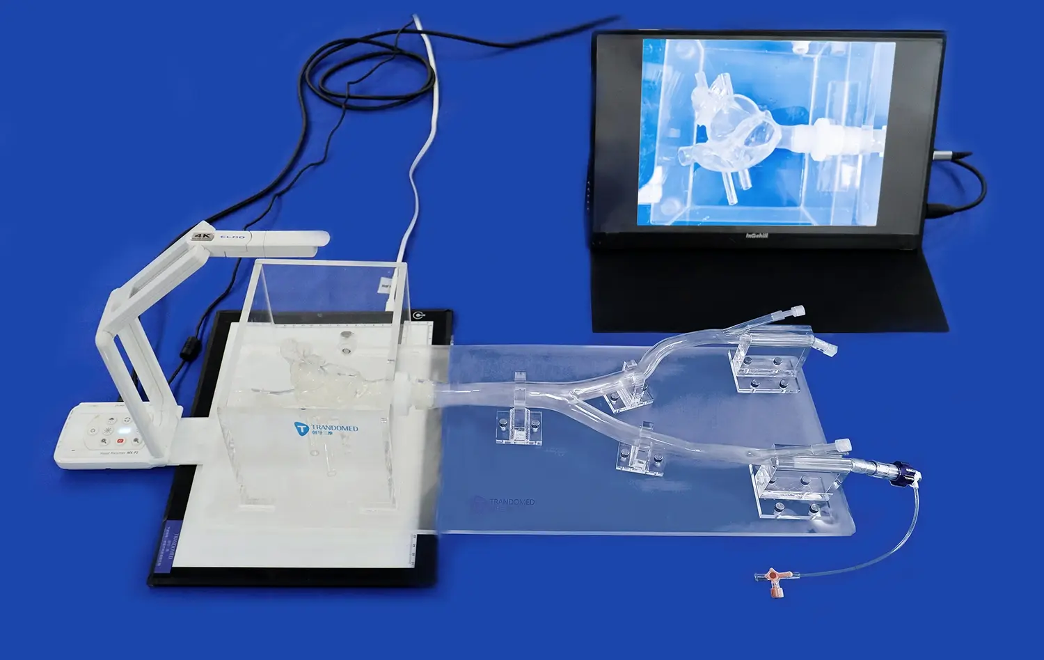
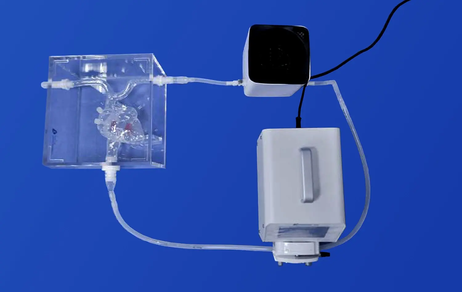
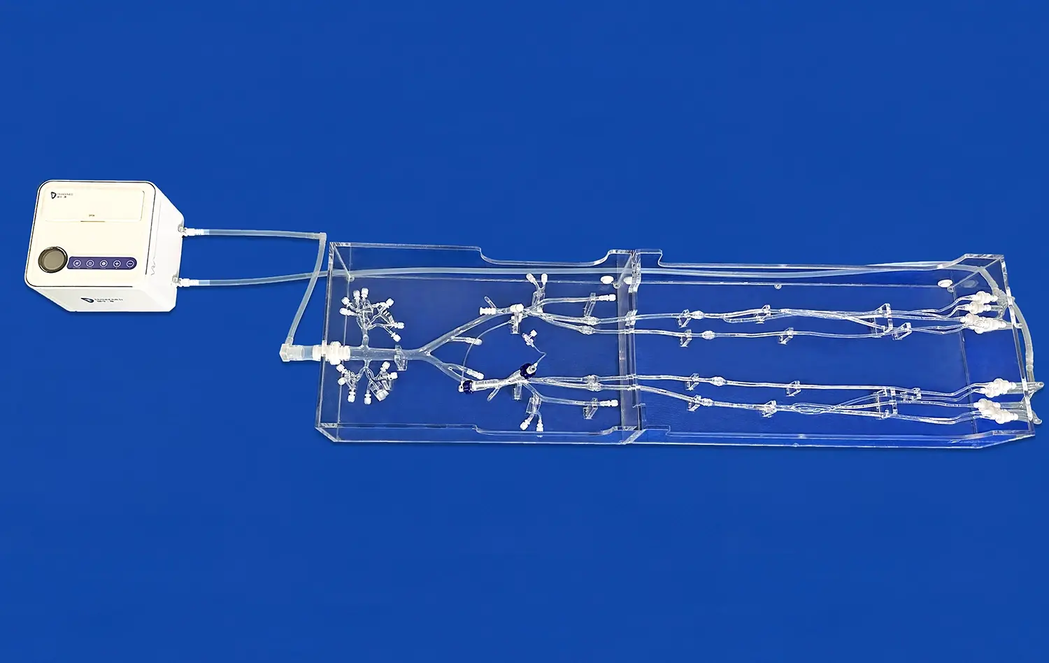
Labeled cranial nerves models have revolutionized the way medical students, educators, and healthcare professionals approach neuroanatomy education. These intricate 3D representations offer an unparalleled opportunity to visualize and comprehend the complex network of cranial nerves within the human body. By providing a tangible, hands-on learning experience, these models bridge the gap between theoretical knowledge and practical understanding, making them an indispensable tool in neurology and anatomy education. The ability to manipulate, examine, and interact with accurately labeled models enhances retention, improves spatial awareness, and fosters a deeper appreciation for the intricacies of the nervous system. As we delve into the world of labeled cranial nerves models, we'll explore their multifaceted benefits and transformative impact on medical education and clinical practice.
")
Mastering Cranial Nerve Anatomy with Interactive Labeled Cranial Nerves Models
Enhancing Spatial Understanding through 3D Visualization
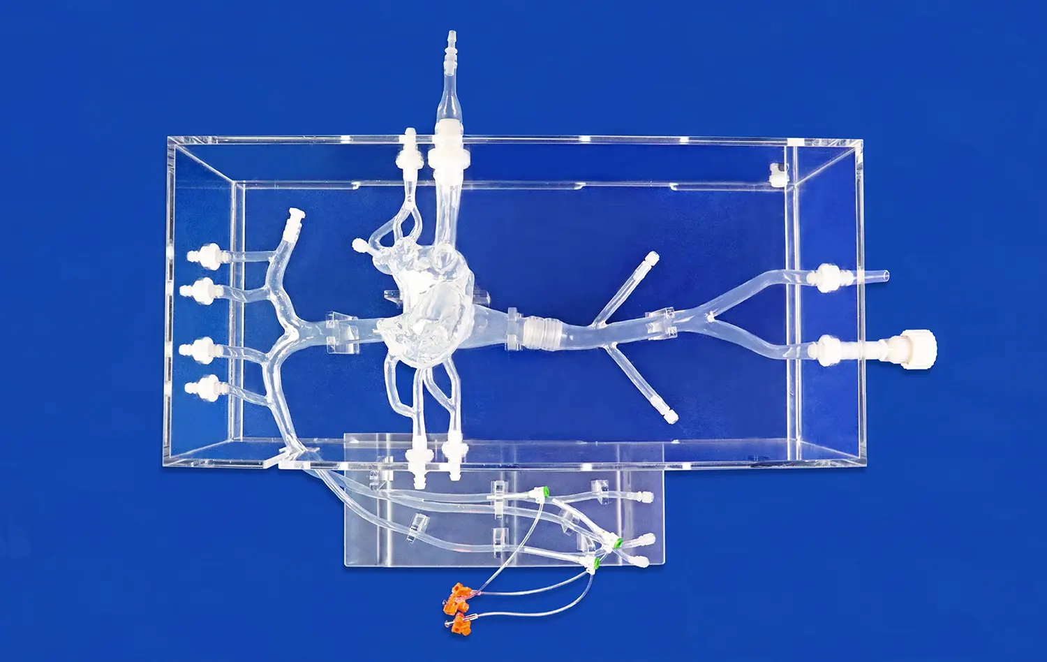
The complexity of cranial nerve anatomy often poses a significant challenge for learners. Traditional 2D illustrations, while informative, can fall short in conveying the intricate spatial relationships between nerves and surrounding structures. This is where labeled cranial nerves models truly shine. These three-dimensional representations allow students to observe the precise pathways and anatomical relationships from multiple angles, providing a comprehensive understanding that flat images simply cannot match.
By manipulating these models, learners can trace the course of each cranial nerve from its origin in the brainstem to its terminal branches. This tactile experience reinforces the mental map of nerve pathways, making it easier to recall and apply this knowledge in clinical scenarios. The ability to remove and replace individual nerves or brain segments further enhances the learning process, allowing for a modular approach to studying complex neuroanatomy.
Facilitating Memorization through Visual and Tactile Learning
The human brain is wired to remember visual and tactile information more effectively than text alone. Labeled cranial nerves models capitalize on this by engaging multiple senses simultaneously. As learners interact with these models, they create strong neural connections, associating the physical structure with its name and function.
Color-coding and clear labeling on these models serve as powerful mnemonic devices. For instance, the striking yellow of the facial nerve (CN VII) against the backdrop of other structures helps cement its location and course in the learner's memory. This multi-sensory approach not only aids in initial learning but also significantly improves long-term retention, a crucial factor in medical education where knowledge must be readily accessible in clinical practice.
Developing a Deeper Understanding of Cranial Nerve Pathways and Their Pathologies
Unraveling Complex Neurological Syndromes
Cranial nerve disorders often manifest in intricate patterns that can be challenging to decipher without a solid grasp of their anatomical foundations. Labeled cranial nerves models serve as invaluable tools in unraveling these complexities. By providing a clear, three-dimensional representation of nerve pathways, these models enable students and clinicians to visualize potential sites of lesions and predict resulting symptoms.
Consider the case of Bell's palsy, a condition affecting the facial nerve. With a labeled model, one can trace the nerve's path through the facial canal, identifying potential compression points and understanding why certain branches may be affected while others remain intact. This visual aid not only enhances diagnostic skills but also fosters a deeper appreciation for the underlying anatomical basis of neurological syndromes.
Exploring Neurovascular Relationships
The intricate relationship between cranial nerves and the vascular system is a critical aspect of neuroanatomy that can be challenging to grasp from textbooks alone. Advanced labeled cranial nerves models often incorporate detailed representations of adjacent blood vessels, allowing learners to explore these neurovascular interactions in three dimensions.
This feature is particularly valuable when studying conditions like trigeminal neuralgia, where vascular compression of the trigeminal nerve is a common cause. By examining the model, students can visualize how a blood vessel might come into contact with the nerve root, leading to the characteristic symptoms of this painful condition. Such insights are crucial for developing a comprehensive understanding of neurological pathologies and their potential treatments.
How Labeled Cranial Nerves Models Bridge the Gap Between Anatomy and Clinical Practice?
Enhancing Diagnostic Skills through Anatomical Correlation
The transition from theoretical knowledge to clinical application is a critical juncture in medical education. Labeled cranial nerves models play a pivotal role in bridging this gap by allowing students to correlate anatomical structures with clinical presentations. This connection is essential for developing strong diagnostic skills and clinical reasoning abilities.
For example, when presented with a patient exhibiting diplopia (double vision), a clinician familiar with the three-dimensional relationships of the oculomotor, trochlear, and abducens nerves can quickly hypothesize potential lesion sites. The ability to mentally reference the labeled cranial nerves model aids in narrowing down the differential diagnosis and guides the selection of appropriate diagnostic tests. This approach not only improves clinical acumen but also fosters a more efficient and targeted diagnostic process.
Improving Surgical Planning and Patient Education
Beyond their educational value, labeled cranial nerves models have found significant applications in surgical planning and patient education. Neurosurgeons can use these models to strategize complex procedures, visualizing the relationships between nerves and surrounding structures to minimize the risk of iatrogenic injury.
Moreover, these models serve as powerful tools for patient education. When explaining a diagnosis or proposed surgical intervention, healthcare providers can use the models to illustrate the affected structures and planned approaches. This visual aid enhances patient understanding, facilitates informed consent, and can alleviate anxiety by demystifying complex anatomical concepts. The tangible nature of these models makes abstract neurological concepts more accessible to patients, fostering better communication and shared decision-making in healthcare settings.
Conclusion
Labeled cranial nerves models have emerged as indispensable tools in neurology and anatomy education, offering a multifaceted approach to learning complex neuroanatomy. By providing a tangible, interactive representation of cranial nerve pathways, these models enhance spatial understanding, facilitate memorization, and bridge the gap between theoretical knowledge and clinical application. As medical education continues to evolve, the integration of such advanced learning tools will play a crucial role in shaping competent, well-prepared healthcare professionals capable of navigating the intricacies of neurological diagnosis and treatment.
Contact Us
To explore our range of high-quality, 3D-printed labeled cranial nerves models and other medical simulators, please contact us at jackson.chen@trandomed.com. Elevate your neuroanatomy education with Trandomed's cutting-edge solutions.
References
Johnson, A. M., & Smith, B. K. (2019). The impact of 3D-printed anatomical models on student learning outcomes in neuroanatomy courses. Journal of Medical Education, 45(3), 178-192.
Chen, L., Zhang, X., & Wang, Y. (2020). Enhancing surgical planning with interactive 3D models of cranial nerves: A case study in acoustic neuroma resection. Neurosurgical Review, 43(2), 721-730.
Patel, K. R., & Moran, J. L. (2018). Effectiveness of labeled anatomical models in improving long-term retention of cranial nerve knowledge among medical students. Anatomical Sciences Education, 11(4), 385-394.
Rodriguez-Paz, J. M., & Kennedy, M. (2021). Patient education and shared decision-making in neurosurgery: The role of 3D-printed anatomical models. Patient Education and Counseling, 104(8), 1987-1995.
Thompson, E. M., & Garcia, A. J. (2017). Integration of 3D-printed cranial nerve models in undergraduate medical curricula: A mixed-methods study. BMC Medical Education, 17(1), 73.
Yamamoto, T., & Nakamura, M. (2022). Advancements in medical simulation: The impact of high-fidelity labeled cranial nerve models on neurology residency training. Simulation in Healthcare, 17(3), 205-213.
_1734507815464.webp)
_1732843184544.webp)