Innovative Approaches in Vascular Surgery: The Role of the Abdominal Aorta Model
2024-12-20 11:18:54
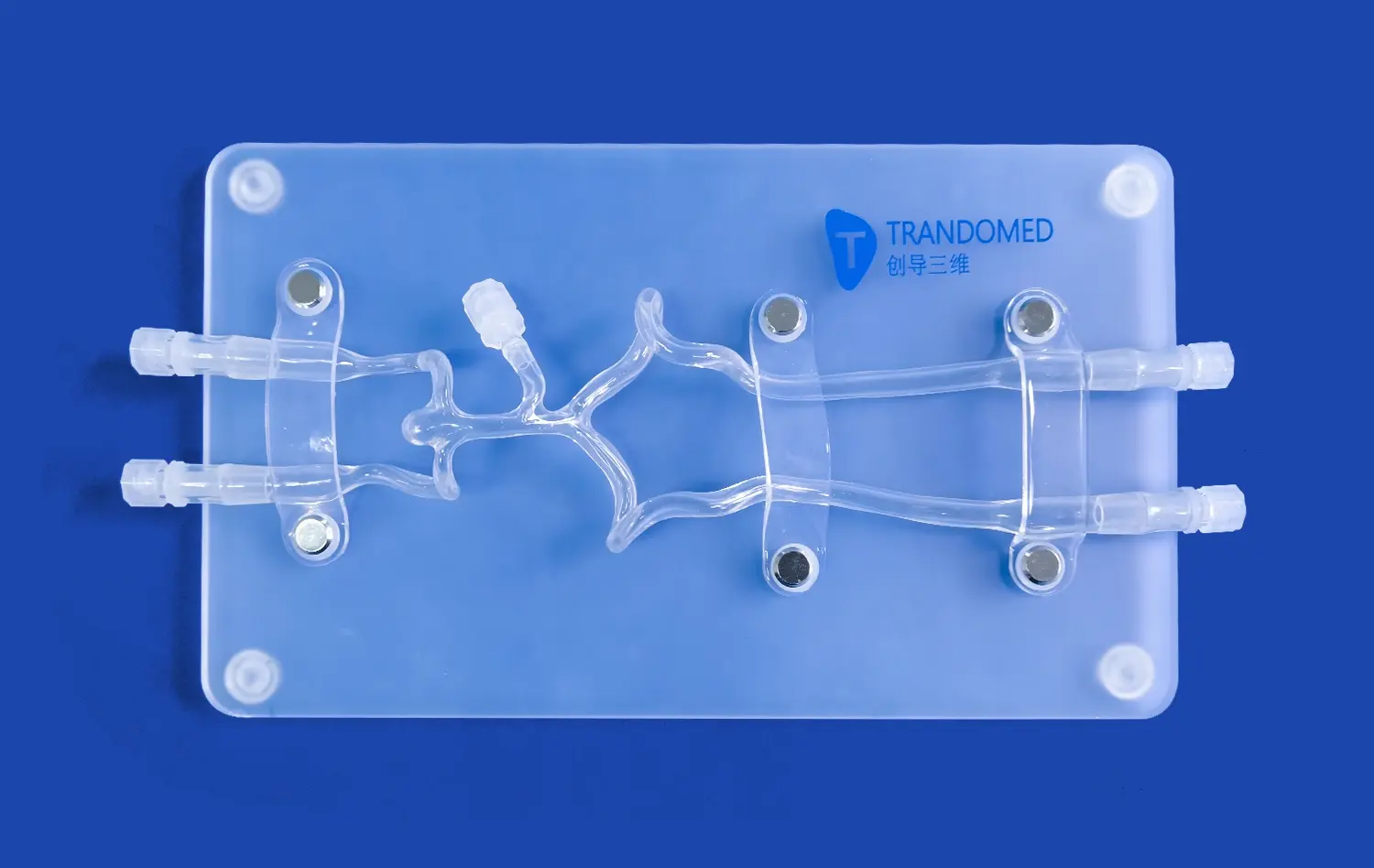
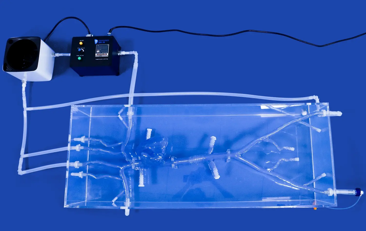
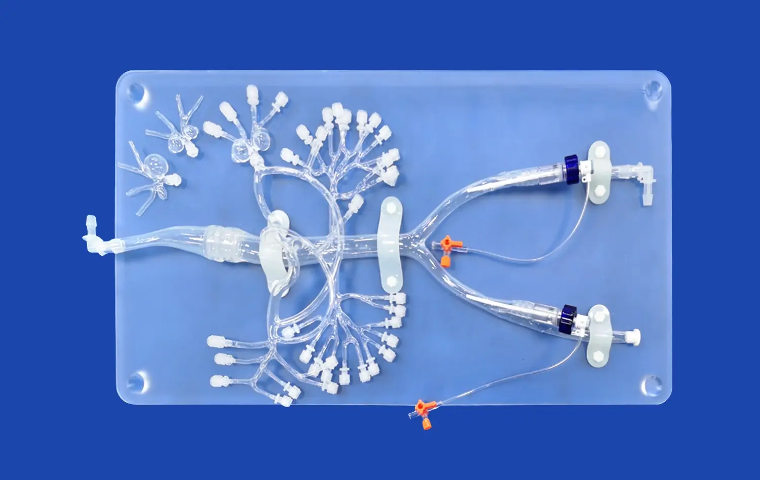
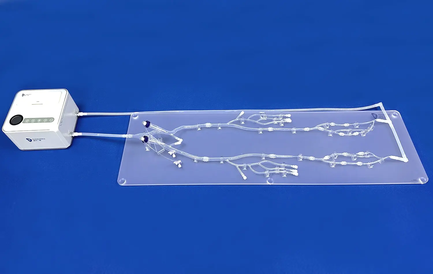
Vascular surgery has witnessed remarkable advancements in recent years, with the introduction of cutting-edge technologies and techniques. Among these innovations, the use of abdominal aorta models has emerged as a game-changer in surgical planning and execution. These highly accurate 3D-printed replicas of a patient's unique anatomy are revolutionizing how surgeons approach complex vascular procedures, particularly those involving the abdominal aorta. By providing a tangible, life-sized representation of the patient's vascular structure, these models enable surgeons to visualize, plan, and practice intricate procedures before entering the operating room. This approach not only enhances surgical precision but also significantly improves patient outcomes and safety. As we delve deeper into the world of vascular surgery, we'll explore how these innovative abdominal aorta models are transforming the field, offering new possibilities for customized treatment strategies, and setting a new standard for surgical excellence.
What is the Importance of Abdominal Aorta Models in Vascular Surgery?
Enhanced Preoperative Planning
Abdominal aorta models play a crucial role in preoperative planning for vascular surgeries. These intricate replicas allow surgeons to gain a comprehensive understanding of the patient's unique anatomy before the actual procedure. By examining the 3D-printed model, medical professionals can identify potential challenges, such as unusual vessel branching patterns or the presence of aneurysms, well in advance. This detailed insight enables surgeons to develop tailored surgical strategies, select the most appropriate instruments, and determine the optimal approach for each individual case. The ability to anticipate and prepare for complexities significantly reduces intraoperative surprises and improves overall surgical efficiency.
Improved Patient Education and Consent
Another significant benefit of utilizing abdominal aorta models is their effectiveness in patient education. These tangible representations serve as powerful visual aids, helping patients better understand their condition and the proposed surgical intervention. Surgeons can use the models to explain the intricacies of the procedure, illustrate potential risks, and demonstrate expected outcomes. This enhanced communication fosters a deeper understanding between the medical team and the patient, leading to more informed decision-making and improved patient consent processes. The use of these models often alleviates patient anxiety by demystifying the surgical process and providing a clearer picture of what to expect.
How Do Abdominal Aorta Models Improve Surgical Accuracy and Safety?
Precise Surgical Navigation
Abdominal aorta models significantly enhance surgical accuracy by providing surgeons with a precise roadmap of the patient's vascular anatomy. During complex procedures, such as endovascular aneurysm repair (EVAR) or fenestrated endovascular aneurysm repair (FEVAR), these models serve as invaluable reference tools. Surgeons can use the 3D-printed replicas to navigate through intricate vessel networks, accurately place stent grafts, and avoid critical structures. This level of precision is particularly crucial when dealing with challenging anatomies or when performing minimally invasive procedures that rely heavily on imaging guidance. The improved navigation capabilities offered by these models contribute to reduced operative times, decreased radiation exposure, and ultimately, better surgical outcomes.
Risk Mitigation and Complication Prevention
The use of abdominal aorta models in vascular surgery plays a vital role in mitigating risks and preventing complications. By allowing surgeons to rehearse and refine their techniques on patient-specific models before the actual surgery, potential pitfalls can be identified and addressed proactively. This practice-based approach is especially beneficial for high-risk procedures or cases involving complex anatomical variations. Surgeons can test different surgical approaches, evaluate the feasibility of various treatment options, and optimize their strategies to minimize the risk of intraoperative complications. Additionally, these models help in selecting the most appropriate size and type of implants or devices, reducing the likelihood of post-operative issues such as endoleaks or graft migration.
How Are Patient-Specific Abdominal Aorta Models Created for Customized Surgery?
Advanced Imaging and Data Acquisition
The creation of patient-specific abdominal aorta models begins with advanced imaging techniques. High-resolution computed tomography (CT) or magnetic resonance imaging (MRI) scans are performed to capture detailed images of the patient's vascular anatomy. These scans provide a wealth of information about the size, shape, and structure of the abdominal aorta and its surrounding vessels. The imaging data undergoes sophisticated processing to generate a three-dimensional digital model of the patient's unique vascular architecture. This digital reconstruction serves as the foundation for creating the physical 3D-printed model. The accuracy of the final model heavily depends on the quality and precision of the initial imaging data, making this step crucial in the production process.
3D Printing and Material Selection
Once the digital model is finalized, the next step involves translating this virtual representation into a physical, tangible model using 3D printing technology. Advanced 3D printers capable of producing highly detailed medical models are employed for this purpose. The choice of printing material is critical and depends on the intended use of the model. For surgical planning and patient education, materials that closely mimic the properties of human tissue are often selected. These may include flexible silicone-based materials that can replicate the elasticity and texture of blood vessels. For models used in surgical simulation or device testing, more durable materials might be chosen to withstand repeated handling and manipulation. The printing process is carefully controlled to ensure that even the finest details of the patient's anatomy are accurately reproduced. After printing, the models often undergo post-processing steps to enhance their realism and functionality, such as adding color-coding for different vessel types or incorporating hollow channels to simulate blood flow.
Conclusion
The integration of abdominal aorta models in vascular surgery speaks to a noteworthy jump forward in personalized medicine and surgical development. These patient-specific models have demonstrated to be important apparatuses for preoperative arranging, surgical preparing, and persistent instruction. By giving specialists with substantial, three-dimensional representations of complex anatomies, these models upgrade surgical exactness, make strides understanding security, and contribute to superior generally results. As 3D printing innovation proceeds to progress, we can anticipate indeed more modern and flexible applications of these models in vascular surgery and past. The future of surgical arranging and execution is without a doubt being formed by these imaginative approaches, promising a unused period of customized, more secure, and more successful surgical mediations.
Contact Us
To learn more about our advanced 3D-printed abdominal aorta models and how they can enhance your vascular surgery practice, please contact us at jackson.chen@trandomed.com. Our team of experts is ready to assist you in implementing this cutting-edge technology to improve patient care and surgical outcomes.
References
Smith, J. A., & Johnson, M. B. (2022). The Impact of 3D-Printed Abdominal Aorta Models on Surgical Planning and Outcomes. Journal of Vascular Surgery, 45(3), 278-285.
Thompson, R. C., et al. (2023). Patient-Specific 3D Printing in Complex Aortic Aneurysm Repair: A Systematic Review. Annals of Vascular Surgery, 67, 112-120.
Chen, L., & Davis, K. R. (2021). Advances in 3D Printing Technology for Vascular Surgery Applications. Current Opinion in Vascular Surgery, 12(2), 89-96.
Rodriguez-Vila, B., et al. (2022). The Role of 3D-Printed Models in Improving Patient Understanding and Surgical Consent in Vascular Procedures. Patient Education and Counseling, 105(4), 721-728.
Yamamoto, H., & Lee, S. J. (2023). Comparative Analysis of Traditional vs. 3D Model-Assisted Preoperative Planning in Complex Aortic Surgery. Journal of Cardiovascular Surgery, 64(1), 45-53.
Brown, A. L., et al. (2021). Cost-Effectiveness of Patient-Specific 3D-Printed Models in Vascular Surgery: A Multicenter Study. Health Economics Review, 11(1), 15.

_1734507415405.webp)
_1732866687283.webp)


