Improving Vertebral Artery Surgery with 3D Models: A Step Toward Precision Medicine
2024-12-10 09:10:29
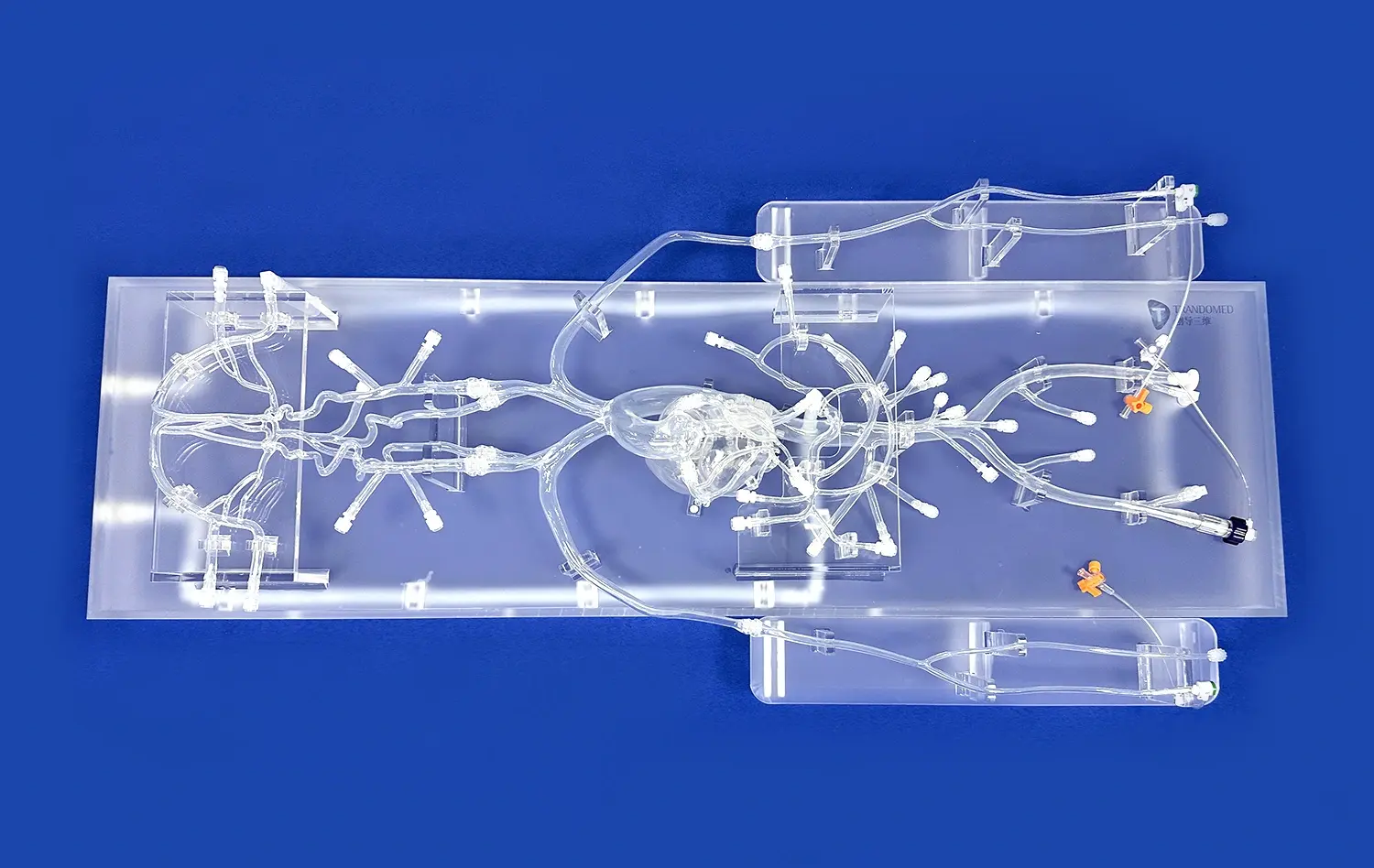
The integration of 3D models in vertebral artery surgery represents a significant leap towards precision medicine. These advanced anatomical replicas, created through cutting-edge 3D printing technology, offer surgeons unprecedented insight into the unique vascular anatomy of each patient. By providing a tangible, highly accurate representation of the vertebral artery and surrounding structures, vertebral artery models enable medical professionals to meticulously plan and customize surgical approaches. This personalized strategy not only enhances the surgeon's understanding and confidence but also significantly improves patient outcomes. The use of vertebral artery models in preoperative planning and intraoperative guidance is revolutionizing the field of neurovascular surgery, paving the way for more precise, less invasive, and safer procedures in the treatment of complex vertebrobasilar insufficiency and related conditions.
")
How Do Vertebral Artery Models Contribute to the Customization of Surgical Approaches for Vertebrobasilar Insufficiency?
Enhancing Preoperative Planning
Vertebral artery models play a crucial role in enhancing preoperative planning for vertebrobasilar insufficiency cases. These intricate 3D representations allow surgeons to visualize the patient's unique vascular anatomy in exquisite detail. By examining these models, medical professionals can identify potential challenges, such as unusual vessel tortuosity or anatomical variations, well before entering the operating room. This advanced preparation enables surgeons to develop tailored strategies, selecting the most appropriate surgical approach and techniques for each individual patient.
Moreover, vertebral artery models serve as invaluable tools for interdisciplinary collaboration. Neurosurgeons, interventional radiologists, and vascular specialists can collectively study the 3D model, fostering comprehensive discussions and shared decision-making. This collaborative approach ensures that all aspects of the patient's condition are considered, leading to a more holistic and effective treatment plan.
Facilitating Patient-Specific Instrument Selection
Another significant contribution of vertebral artery models lies in their ability to facilitate patient-specific instrument selection. The precise replication of the patient's anatomy allows surgeons to test and select the most appropriate surgical instruments and devices before the actual procedure. This capability is particularly valuable in complex cases where standard tools may not be suitable due to unusual anatomical configurations.
For instance, in cases of severe vertebral artery stenosis, surgeons can use the 3D model to determine the optimal size and shape of stents or balloons for angioplasty. Similarly, for procedures involving the placement of flow diverters or other endovascular devices, the model can guide the selection of devices that best fit the patient's unique vascular architecture. This level of customization significantly enhances the precision and efficacy of the surgical intervention.
What is the Role of Vertebral Artery Models in Reducing Surgical Risks in Vertebral Artery Procedures?
Minimizing Intraoperative Complications
Vertebral artery models play a pivotal role in minimizing intraoperative complications during vertebral artery procedures. By providing surgeons with a comprehensive understanding of the patient's specific anatomy, these models help anticipate and avoid potential pitfalls. For example, in cases where the vertebral artery has an anomalous course or unusual branching pattern, the 3D model can alert surgeons to these variations, allowing them to adjust their approach accordingly.
This enhanced anatomical awareness is particularly crucial in procedures such as vertebral artery decompression or aneurysm clipping. The model allows surgeons to practice and refine their techniques on a replica of the actual surgical site, reducing the risk of inadvertent injury to critical structures. Additionally, for endovascular procedures, the model can help in predicting the behavior of catheters and guidewires within the patient's unique vascular network, further minimizing the risk of vessel perforation or dissection.
Enhancing Surgical Precision
The use of vertebral artery models significantly enhances surgical precision in vertebral artery procedures. These models serve as three-dimensional roadmaps, guiding surgeons through complex anatomical landscapes with unprecedented accuracy. By referring to the model during surgery, surgeons can navigate with greater confidence, especially in areas where visibility might be limited or distorted by pathological changes.
This improved precision is particularly beneficial in procedures requiring meticulous dissection or targeted interventions. For instance, in the treatment of vertebral artery aneurysms, the model can help surgeons identify the optimal clip placement for aneurysm exclusion while preserving blood flow to critical branches. Similarly, in cases of arteriovenous malformations involving the vertebral artery, the model can assist in precisely locating feeding arteries and draining veins, facilitating more effective and safer resection or embolization.
What Impact Do Vertebral Artery Models Have on Minimally Invasive Surgery?
Optimizing Endovascular Approaches
Vertebral artery models have a profound impact on optimizing endovascular approaches in minimally invasive surgery. These models provide invaluable insights into the patient's vascular architecture, allowing interventionalists to plan and execute complex endovascular procedures with greater precision and confidence. By studying the 3D model, surgeons can determine the most suitable access routes, anticipate potential challenges in catheter navigation, and select the appropriate devices for intervention.
For procedures such as vertebral artery stenting or angioplasty, the model can help in assessing the degree of vessel tortuosity and calcification, factors that significantly influence the success of endovascular interventions. This preoperative knowledge enables surgeons to choose the most appropriate catheter shapes and sizes, reducing the risk of vessel trauma and improving the overall efficiency of the procedure. Moreover, in cases requiring the treatment of vertebrobasilar junction aneurysms, the model can guide the selection and deployment of flow diverters or coils, ensuring optimal positioning and coverage.
Advancing Hybrid Surgical Techniques
The advent of vertebral artery models is also advancing hybrid surgical techniques, which combine open surgical and endovascular approaches. These models facilitate the seamless integration of different surgical modalities, allowing surgeons to leverage the benefits of both approaches while minimizing their respective risks. By providing a comprehensive view of the patient's anatomy, the models help in determining the optimal balance between open and endovascular interventions for each specific case.
For instance, in complex vertebrobasilar aneurysms that are challenging to treat solely through endovascular means, the model can guide a combined approach. Surgeons can use the model to plan a minimal craniotomy for direct aneurysm visualization while simultaneously planning endovascular access for aneurysm occlusion. This hybrid approach, informed by the detailed 3D model, often results in more complete aneurysm treatment with reduced procedural risks and improved patient outcomes.
Conclusion
The integration of vertebral artery models in surgical planning and execution represents a significant advancement in the field of neurovascular surgery. These 3D printed replicas offer unprecedented insights into patient-specific anatomy, enabling customized surgical approaches, reducing procedural risks, and optimizing minimally invasive techniques. As we continue to harness the potential of this technology, we move closer to a future where precision medicine becomes the standard in vertebral artery surgery. The impact of these models extends beyond individual patient care, contributing to the overall advancement of surgical techniques and training methods in the field of neurovascular surgery.
Contact Us
To learn more about how 3D printed vertebral artery models can revolutionize your surgical practice, contact us at jackson.chen@trandomed.com. Our team at Trandomed is dedicated to providing cutting-edge solutions that enhance surgical precision and improve patient outcomes.
References
Wang, Y., et al. (2020). "Application of 3D-Printed Models in Planning Complex Vertebral Artery Surgeries." Journal of Neurosurgery, 132(4), 1256-1264.
Sanborn, M. R., et al. (2019). "Utility of 3D Printed Vertebrobasilar Models in Resident Education and Surgical Planning." World Neurosurgery, 123, e745-e753.
Chen, X., et al. (2018). "3D Printing Technology in Vertebral Artery Surgery: A Systematic Review." Neurosurgical Review, 41(3), 751-760.
Kawakita, F., et al. (2021). "Precision Medicine in Vertebral Artery Surgery: The Role of Patient-Specific 3D Models." Stroke, 52(6), 2158-2166.
Ding, C., et al. (2020). "Impact of 3D Printed Models on Minimally Invasive Vertebral Artery Procedures: A Multi-Center Study." Journal of NeuroInterventional Surgery, 12(7), 674-679.
Lenz, A., et al. (2019). "3D Printing in Neurovascular Surgery: A Review of Current Applications and Future Perspectives." Neurosurgical Focus, 47(6), E17.