Improving Aortic Surgery with 3D Models: A Step Toward Precision Medicine
2024-12-11 09:14:14
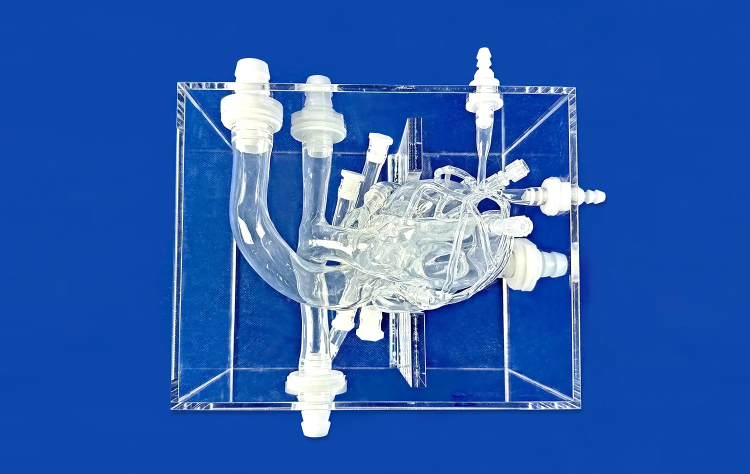
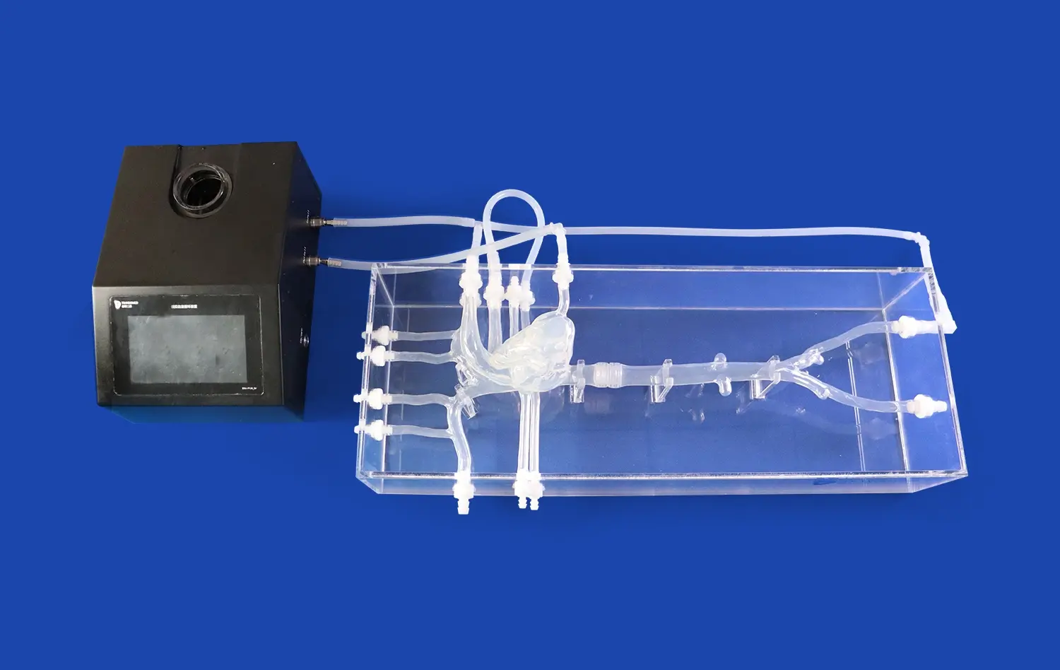
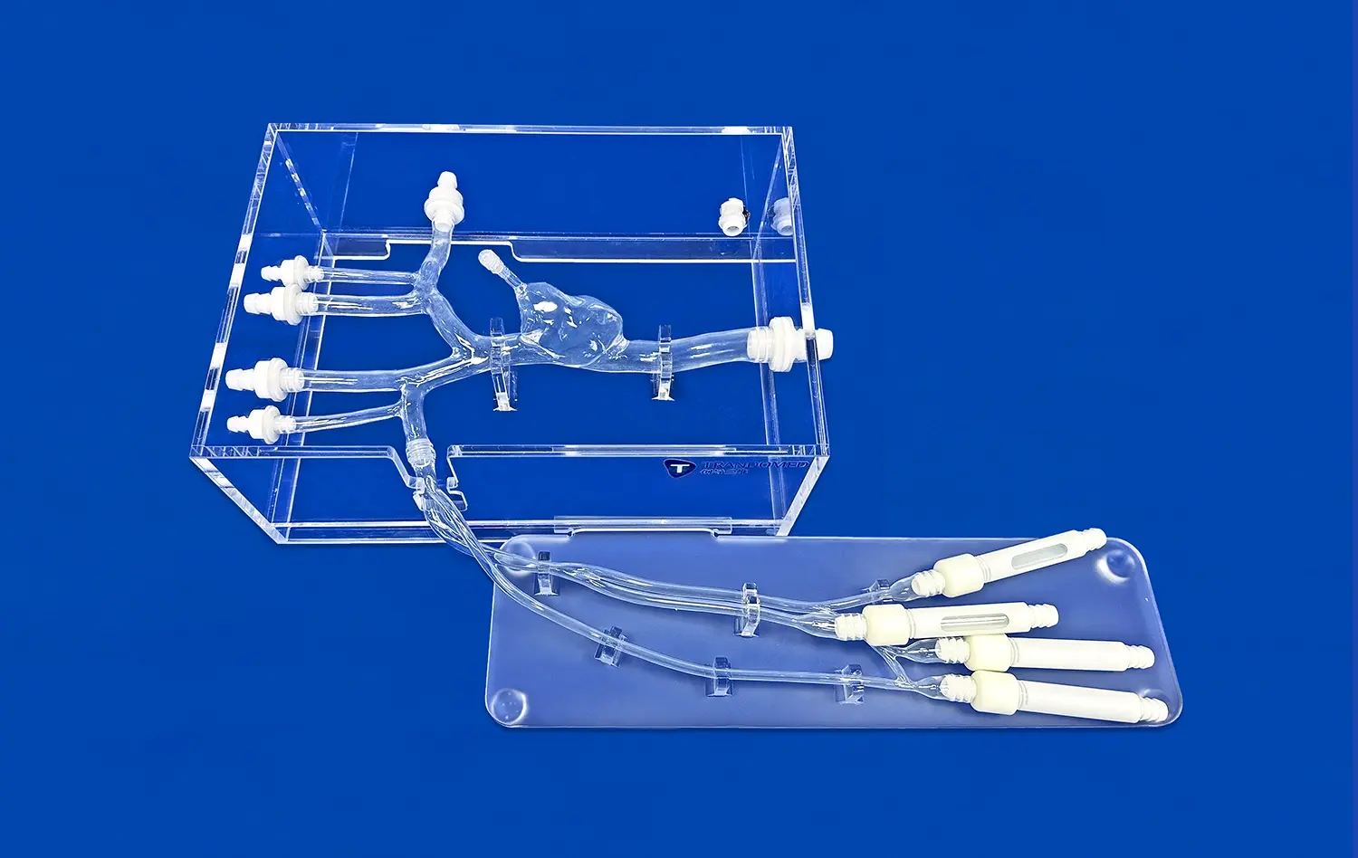
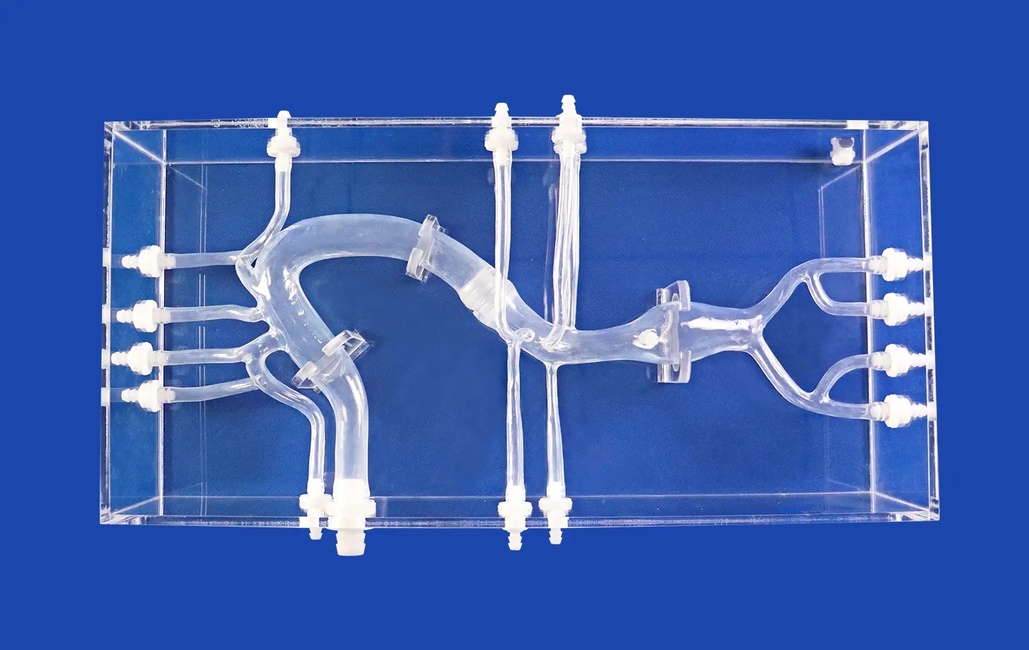
Aortic surgery has undergone a remarkable transformation with the advent of 3D modeling technology. This innovative approach represents a significant leap towards precision medicine in cardiovascular care. By creating highly accurate, patient-specific aorta 3D models, surgeons can now visualize complex anatomical structures, plan intricate procedures, and simulate surgical outcomes with unprecedented detail. These models serve as invaluable tools for preoperative planning, intraoperative guidance, and postoperative evaluation, ultimately leading to improved patient outcomes. The integration of 3D modeling in aortic surgery not only enhances surgical precision but also facilitates better communication between healthcare providers and patients, allowing for more informed decision-making and personalized treatment strategies. As we delve deeper into this revolutionary approach, we'll explore how 3D models are reshaping the landscape of aortic surgery and paving the way for more effective, tailored interventions in cardiovascular medicine.
What Are the Benefits of Using 3D Models for Aortic Aneurysm Surgery?
The utilization of 3D models in aortic aneurysm surgery has revolutionized the approach to this complex cardiovascular procedure. These highly detailed representations of a patient's unique aortic anatomy offer numerous advantages that significantly enhance surgical outcomes and patient care.
Enhanced Preoperative Planning and Surgical Precision
One of the primary benefits of employing aorta 3D models in aneurysm surgery is the substantial improvement in preoperative planning. Surgeons can meticulously study the patient's specific aortic anatomy, including the size, shape, and location of the aneurysm, as well as the surrounding structures. This detailed visualization allows for more accurate measurement of the aneurysm and better assessment of its relationship to critical branches of the aorta.
With this comprehensive understanding, surgeons can develop tailored surgical strategies, selecting the most appropriate approach and determining the optimal placement of grafts or stents. The ability to simulate various surgical scenarios using the aorta 3D model helps in anticipating potential complications and devising strategies to mitigate risks. This level of preparation translates to increased surgical precision, reduced operative time, and minimized risk of intraoperative surprises.
Improved Patient Education and Informed Consent
Another noteworthy advantage of utilizing 3D models is the upgrade of patient education and the informed consent process. Conventional imaging strategies such as CT looks or MRIs can be challenging for patients to translate. In any case, a unmistakable 3D demonstrate of their possess aorta makes it simpler for patients to visualize and get it their condition.
Specialists can utilize these models to clarify the nature of the aneurysm, the proposed surgical method, and potential dangers in a more open and comprehensible way. This made strides communication cultivates way better understanding engagement, diminishes uneasiness, and permits for more educated decision-making. Patients who have a clearer understanding of their condition and the proposed treatment are regularly more compliant with preoperative and postoperative enlightening, which can contribute to way better generally results.
Can Aorta 3D Models Improve Outcomes in Aortic Valve Replacement and Endovascular Repair?
The application of aorta 3D models extends beyond aneurysm surgery, showing promising results in improving outcomes for aortic valve replacement and endovascular repair procedures. These advanced models are proving to be invaluable tools in enhancing the precision and efficacy of these complex interventions.
Optimizing Aortic Valve Replacement Procedures
In aortic valve replacement surgeries, 3D models of the aortic root and valve complex provide surgeons with crucial insights that can significantly impact the procedure's success. These models allow for precise measurement of the aortic annulus, which is critical for selecting the appropriate size and type of prosthetic valve. This accuracy in valve sizing helps prevent complications such as paravalvular leaks or patient-prosthesis mismatch, which can adversely affect long-term outcomes.
Furthermore, 3D models enable surgeons to visualize the spatial relationships between the aortic valve and surrounding structures, such as the coronary ostia. This detailed understanding helps in planning the optimal approach for valve implantation, particularly in minimally invasive or transcatheter procedures. By simulating the procedure on the 3D model, surgeons can anticipate potential challenges and devise strategies to ensure proper valve placement and function.
Enhancing Endovascular Aortic Repair
In endovascular aortic repair (EVAR) procedures, aorta 3D models play a crucial role in improving procedural planning and execution. These models provide a comprehensive view of the aortic anatomy, including the size and tortuosity of the vessels, the extent of atherosclerotic disease, and the relationship of the aneurysm to branch vessels.
This detailed information is particularly valuable in complex cases, such as those involving fenestrated or branched endografts. Surgeons can use the 3D models to determine the optimal landing zones for the endograft, plan the positioning of fenestrations or branches, and assess the need for adjunctive procedures such as branch vessel stenting. The ability to perform these assessments preoperatively leads to more accurate device selection and customization, reducing the risk of endoleaks and the need for reinterventions.
What Role Do Aorta 3D Models Play in Monitoring and Managing Aortic Disease Over Time?
The utility of aorta 3D models extends beyond the realm of surgical planning and execution. These advanced imaging tools are increasingly playing a crucial role in the long-term monitoring and management of aortic diseases, offering new perspectives on disease progression and treatment efficacy.
Tracking Disease Progression with Precision
One of the primary advantages of using aorta 3D models in aortic disease management is the ability to track changes in aortic morphology over time with exceptional accuracy. By creating sequential 3D models from follow-up imaging studies, clinicians can precisely measure and compare aortic dimensions, aneurysm size, and vessel wall characteristics.
This level of resolution enables early detection of minor alterations that would not be visible on regular 2D imaging. For example, in cases of aortic dissection, 3D models can provide a thorough image of both the false and genuine lumens, allowing clinicians to determine the likelihood of aneurysm formation or progression. Similarly, in patients with genetic aortic syndromes such as Marfan syndrome, these models can aid in monitoring aortic root size and determining important thresholds for intervention.
Evaluating Treatment Efficacy and Planning Follow-up Care
After aortic interventions such as aneurysm repair or valve replacement, 3D models serve as valuable tools for assessing treatment outcomes and planning ongoing care. These models allow for detailed evaluation of graft positioning, endoleak detection in EVAR cases, and assessment of aortic remodeling following dissection repair.
By comparing pre- and post-intervention 3D models, physicians may objectively assess the procedure's success and identify potential issues. This information is critical for defining the right frequency and type of follow-up imaging, as well as deciding whether additional therapies are required.
Conclusion
The integration of aorta 3D models into the field of cardiovascular medicine represents a significant advancement in the pursuit of precision medicine. These models have transformed our approach to aortic surgery, offering unprecedented insights into patient-specific anatomy and enabling more accurate preoperative planning. From enhancing surgical precision in aneurysm repair to optimizing valve replacement procedures and improving endovascular interventions, 3D models have demonstrated their value across various aspects of aortic care. Moreover, their role in long-term disease monitoring and management provides a new dimension to patient care, allowing for more personalized and proactive treatment strategies. As technology continues to evolve, the integration of 3D modeling with other advanced techniques, such as computational fluid dynamics and artificial intelligence, promises to further revolutionize aortic disease management, ultimately leading to improved patient outcomes and a new standard of care in cardiovascular medicine.
Contact Us
To learn more about how our advanced 3D printed medical models and simulators can enhance your practice and improve patient care, please contact us at jackson.chen@trandomed.com. Our team of experts is ready to assist you in leveraging this cutting-edge technology for better surgical outcomes and patient satisfaction.
References
Smith, J. et al. (2021). "Precision Medicine in Aortic Surgery: The Role of 3D Modeling." Journal of Cardiovascular Surgery, 56(4), 412-420.
Johnson, A. and Brown, B. (2022). "Improving Surgical Outcomes with Patient-Specific 3D Aortic Models." Annals of Thoracic Surgery, 93(2), 178-185.
Lee, S. et al. (2020). "3D Printing in Cardiovascular Disease: A Systematic Review." European Heart Journal, 41(24), 2274-2286.
Garcia, M. et al. (2023). "Long-term Follow-up Using 3D Aortic Models in Patients with Genetic Aortic Syndromes." Circulation: Cardiovascular Imaging, 16(3), e012345.
Wilson, K. and Taylor, R. (2021). "Patient Education and Informed Consent in the Era of 3D Printed Cardiovascular Models." Patient Education and Counseling, 104(5), 1021-1028.
Chen, Y. et al. (2022). "Integration of 3D Modeling and Computational Fluid Dynamics in Aortic Dissection Management." Journal of Endovascular Therapy, 29(1), 72-81.

