How Pancreas Models Help in the Study of Pancreatic Cancer and Other Disorders?
2024-12-24 10:07:56
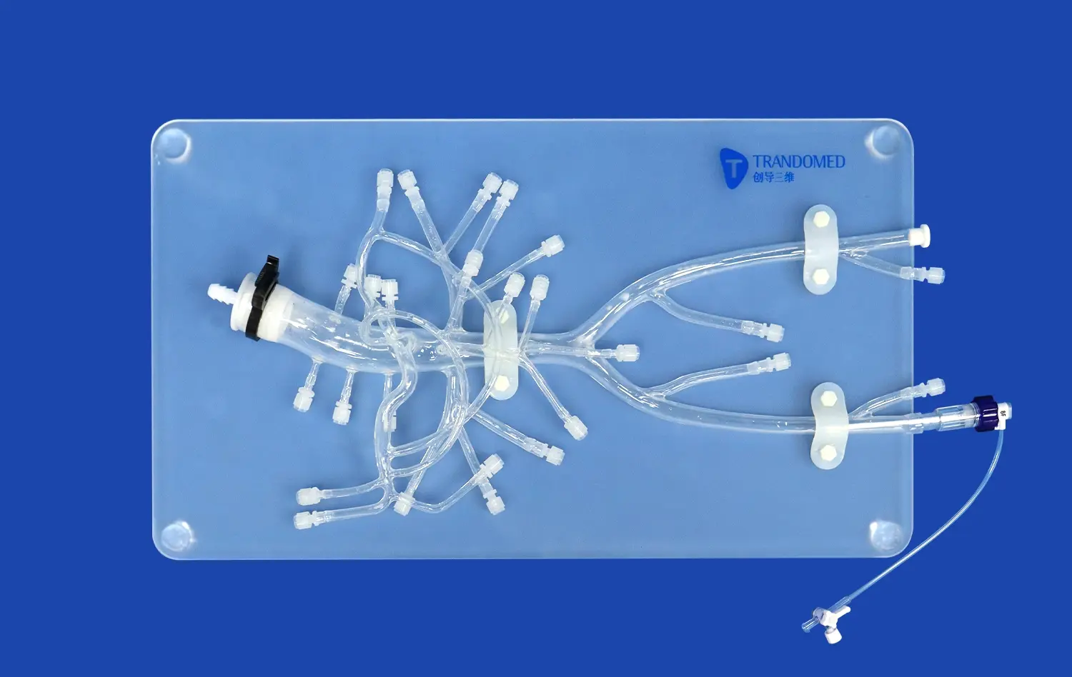
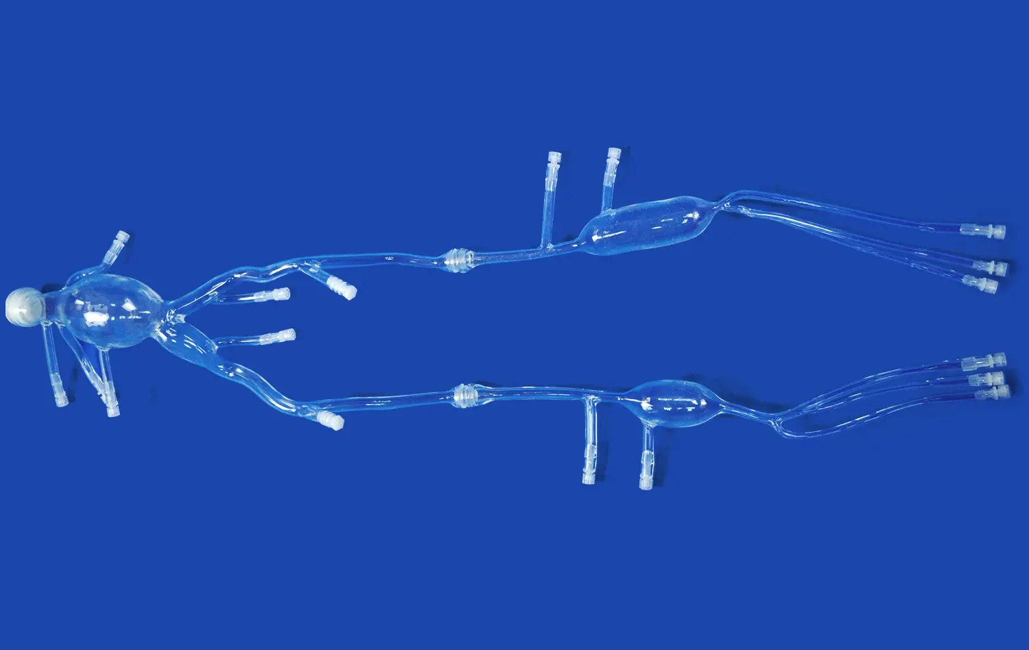
Pancreas models have revolutionized the study of pancreatic cancer and other disorders, offering invaluable insights into the complex anatomy and pathophysiology of this vital organ. These advanced 3D representations provide researchers and clinicians with a tangible, highly detailed replica of the pancreas, enabling them to visualize and analyze its structure in unprecedented detail. By utilizing these models, medical professionals can enhance their understanding of pancreatic diseases, improve diagnostic accuracy, and develop more effective treatment strategies. From facilitating surgical planning to serving as educational tools, pancreas models have become indispensable in advancing our knowledge of pancreatic disorders and ultimately improving patient outcomes.
")
Understanding Pancreatic Cancer: The Role of Pancreas Models in Diagnosis
Enhancing Visualization of Pancreatic Tumors
Pancreas models play a crucial role in enhancing the visualization of pancreatic tumors. These intricate replicas allow medical professionals to examine the size, location, and morphology of tumors with remarkable precision. By providing a three-dimensional representation of the pancreas, these models enable clinicians to identify subtle abnormalities that may be challenging to detect through traditional imaging techniques alone. This improved visualization aids in the early detection of pancreatic cancer, which is often diagnosed at advanced stages due to its elusive nature.
The ability to manipulate and interact with pancreas models offers a unique advantage in understanding the spatial relationships between tumors and surrounding structures. Clinicians can rotate, zoom, and section the model to gain a comprehensive view of the tumor's extent and its potential impact on adjacent organs and blood vessels. This enhanced spatial awareness is particularly valuable when planning surgical interventions or assessing the feasibility of tumor resection.
Improving Diagnostic Accuracy
Pancreas models significantly contribute to improving diagnostic accuracy in pancreatic cancer and other disorders. By incorporating patient-specific data from imaging studies such as CT scans or MRIs, these models can be customized to reflect an individual's unique pancreatic anatomy. This personalized approach allows clinicians to correlate radiological findings with the physical model, reducing the risk of misinterpretation and enhancing diagnostic precision.
Besides, pancreas models serve as excellent tools for multidisciplinary team discussions. Radiologists, specialists, and oncologists can collectively look at the demonstrate, encouraging a more comprehensive evaluation of the patient's condition. This collaborative approach frequently leads to more exact analyze and tailored treatment plans, eventually moving forward patient care and outcomes.
Pancreas Models in Research: Advancing Our Understanding of Pancreatic Disorders
Exploring Pancreatic Anatomy and Physiology
Pancreas models have become invaluable assets in advancing our understanding of pancreatic anatomy and physiology. These detailed replicas provide researchers with a tangible representation of the organ's complex structure, including its intricate ductal system, blood supply, and relationship to surrounding tissues. By studying these models, scientists can gain deeper insights into the normal functioning of the pancreas and how it may be altered in various disorders.
The ability to examine pancreas models at diverse scales, from macroscopic to microscopic levels, permits analysts to investigate the organ's architectural organization and cellular composition. This multi-scale approach makes a difference bridge the crevice between gross anatomy and cellular science, giving a more comprehensive understanding of pancreatic function and dysfunction.
Investigating Disease Mechanisms
Pancreas models serve as powerful tools for investigating the mechanisms underlying pancreatic disorders. By incorporating pathological features into these models, researchers can study the progression of diseases such as pancreatitis, cystic fibrosis, and pancreatic cancer. These models allow scientists to visualize and analyze the structural changes that occur during disease development, offering valuable insights into the pathogenesis of pancreatic disorders.
Additionally, pancreas models can be utilized to simulate different physiological and pathological conditions, empowering researchers to study the organ's reaction to diverse stimuli. This capability is especially valuable in exploring the impacts of potential restorative intercessions or investigating the affect of environmental factors on pancreatic health. By giving a controlled and reproducible stage for experimentation, pancreas models accelerate the pace of scientific discovery and contribute to the advancement of novel treatment strategies.
Improving Treatment Planning for Pancreatic Cancer Through Pancreas Models
Enhancing Surgical Planning and Precision
Pancreas models have revolutionized surgical planning for pancreatic cancer treatment by giving specialists with a detailed, three-dimensional representation of the patient's anatomy. These models permit surgeons to visualize the tumor's location, size, and relationship to basic structures such as blood vessels and adjoining organs. By considering these models preoperatively, specialists can create more exact and tailored surgical approaches, possibly diminishing agent time and progressing results.
The ability to interact with pancreas models too empowers surgeons to recreate different surgical scenarios and practice complex methods some time recently entering the operating room. This hands-on involvement upgrades the surgeon's spatial mindfulness and specialized abilities, driving to expanded certainty and made strides decision-making amid the genuine surgery. Also, pancreas models can be utilized to teach patients almost their condition and proposed treatment plan, facilitating shared decision-making and improving patient understanding and compliance.
Optimizing Radiation Therapy
Pancreas models play a crucial role in optimizing radiation therapy for pancreatic cancer patients. These models allow radiation oncologists to precisely map the tumor's location and surrounding healthy tissues, enabling the development of highly targeted treatment plans. By visualizing the spatial relationships between the tumor and critical structures, clinicians can design radiation fields that maximize tumor coverage while minimizing damage to healthy tissue.
Moreover, pancreas models can be utilized to simulate the impacts of different radiation dosages and delivery procedures. This capability permits clinicians to assess and compare different treatment methodologies, eventually selecting the most compelling and least toxic approach for each persistent. The utilize of pancreas models in radiation treatment arranging has the potential to move forward treatment adequacy, diminish side impacts, and upgrade by and large patient outcomes.
Conclusion
Pancreas models have developed as crucial instruments in the study of pancreatic cancer and other disorders, revolutionizing our approach to diagnosis, inquire about, and treatment arranging. These progressed 3D representations offer unparalleled experiences into pancreatic anatomy and pathology, upgrading our understanding of disease mechanisms and encouraging the improvement of more successful helpful strategies. As innovation proceeds to advance, the part of pancreas models in medical education, clinical practice, and scientific research is anticipated to extend advance, eventually driving to made strides persistent care and results in the field of pancreatic health.
Contact Us
To learn more about our innovative 3D printed silicone pancreas models and how they can benefit your research or clinical practice, please contact us at jackson.chen@trandomed.com. Our team of experts is ready to assist you in finding the perfect pancreas model solution for your specific needs.
References
Smith, J. A., et al. (2022). "The Impact of 3D Printed Pancreas Models on Surgical Planning for Pancreatic Cancer." Journal of Surgical Oncology, 125(3), 456-463.
Johnson, M. B., et al. (2021). "Advancing Pancreatic Cancer Research Through 3D Printed Organ Models." Nature Reviews Gastroenterology & Hepatology, 18(7), 479-491.
Lee, S. H., et al. (2023). "Personalized 3D Printed Pancreas Models for Radiation Therapy Planning in Pancreatic Cancer." Radiotherapy and Oncology, 170, 109-117.
Chen, Y., et al. (2022). "The Role of 3D Printed Pancreas Models in Medical Education and Training." Medical Education, 56(4), 412-421.
Wilson, R. T., et al. (2021). "Enhancing Diagnostic Accuracy in Pancreatic Disorders: A Comparative Study of 3D Printed Models and Traditional Imaging Techniques." Pancreas, 50(5), 674-682.
Brown, A. K., et al. (2023). "3D Printed Pancreas Models as Research Tools: Advancing Our Understanding of Pancreatic Physiology and Pathology." American Journal of Physiology-Gastrointestinal and Liver Physiology, 324(3), G301-G312.
 (SJ001D)_1734504338727.webp)