How Detachable Coronary Models Assist in Understanding Heart Disease Pathology?
2024-11-27 16:11:23
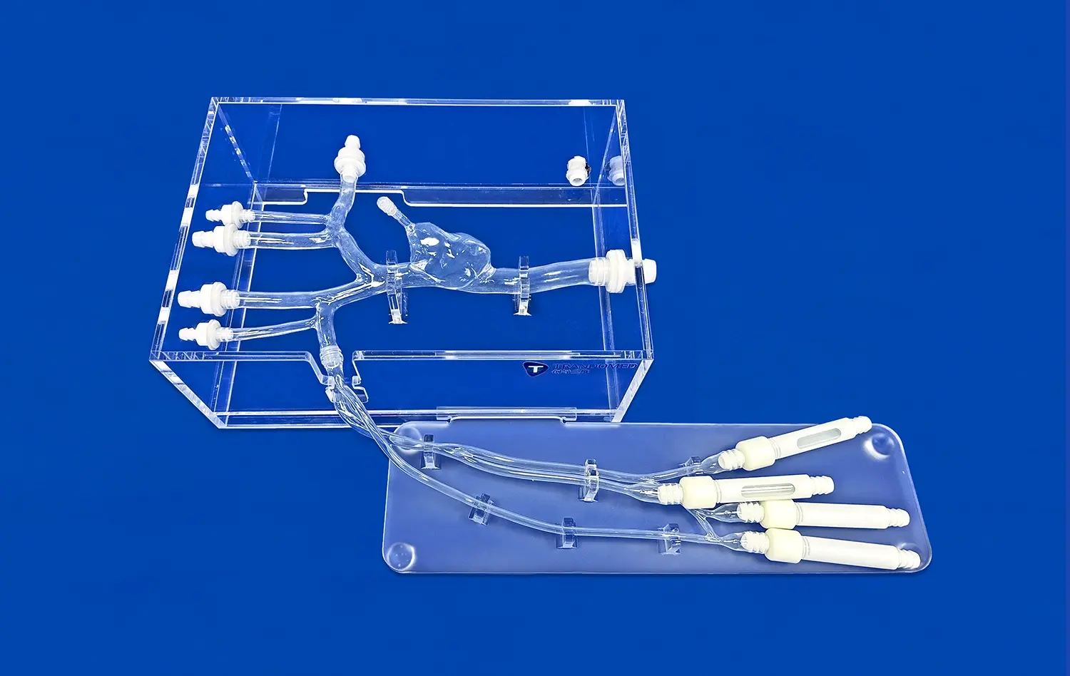
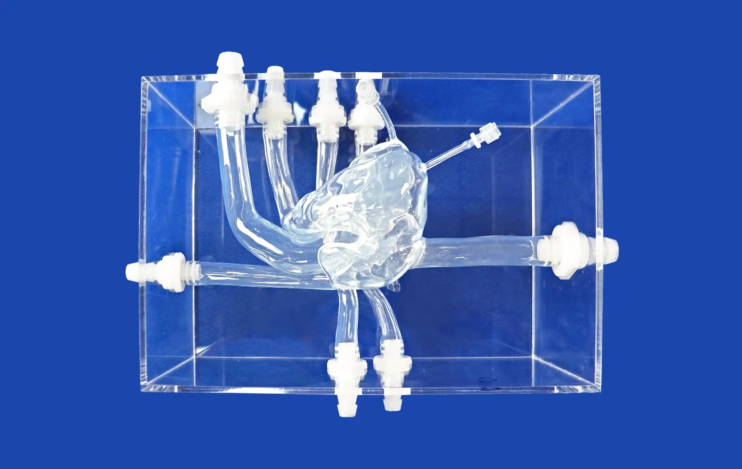
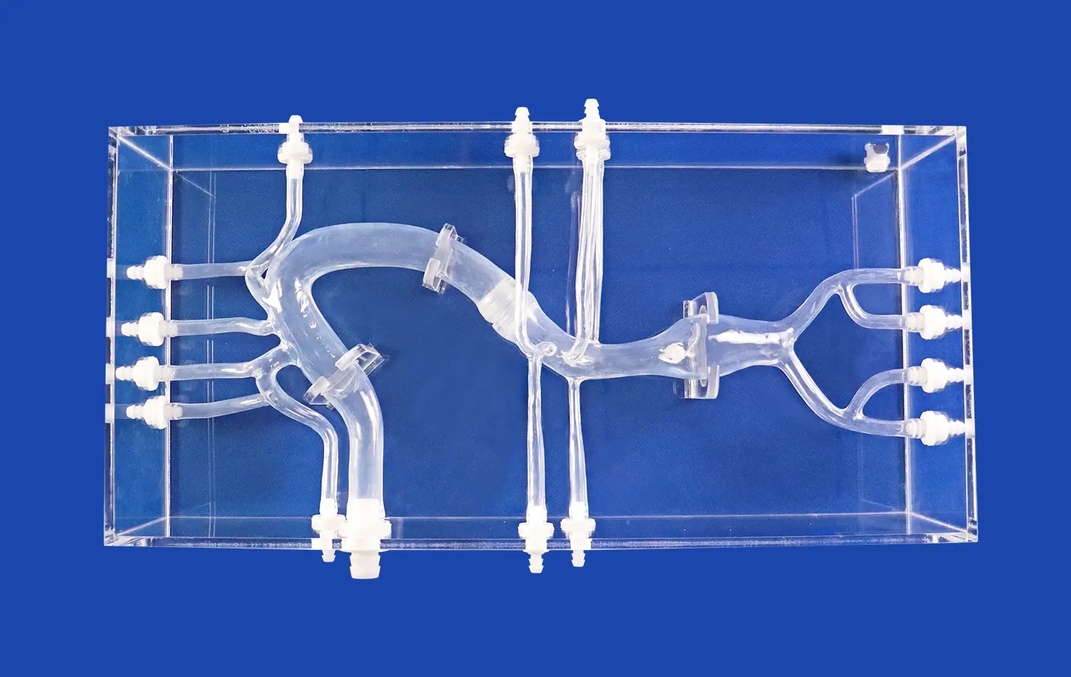
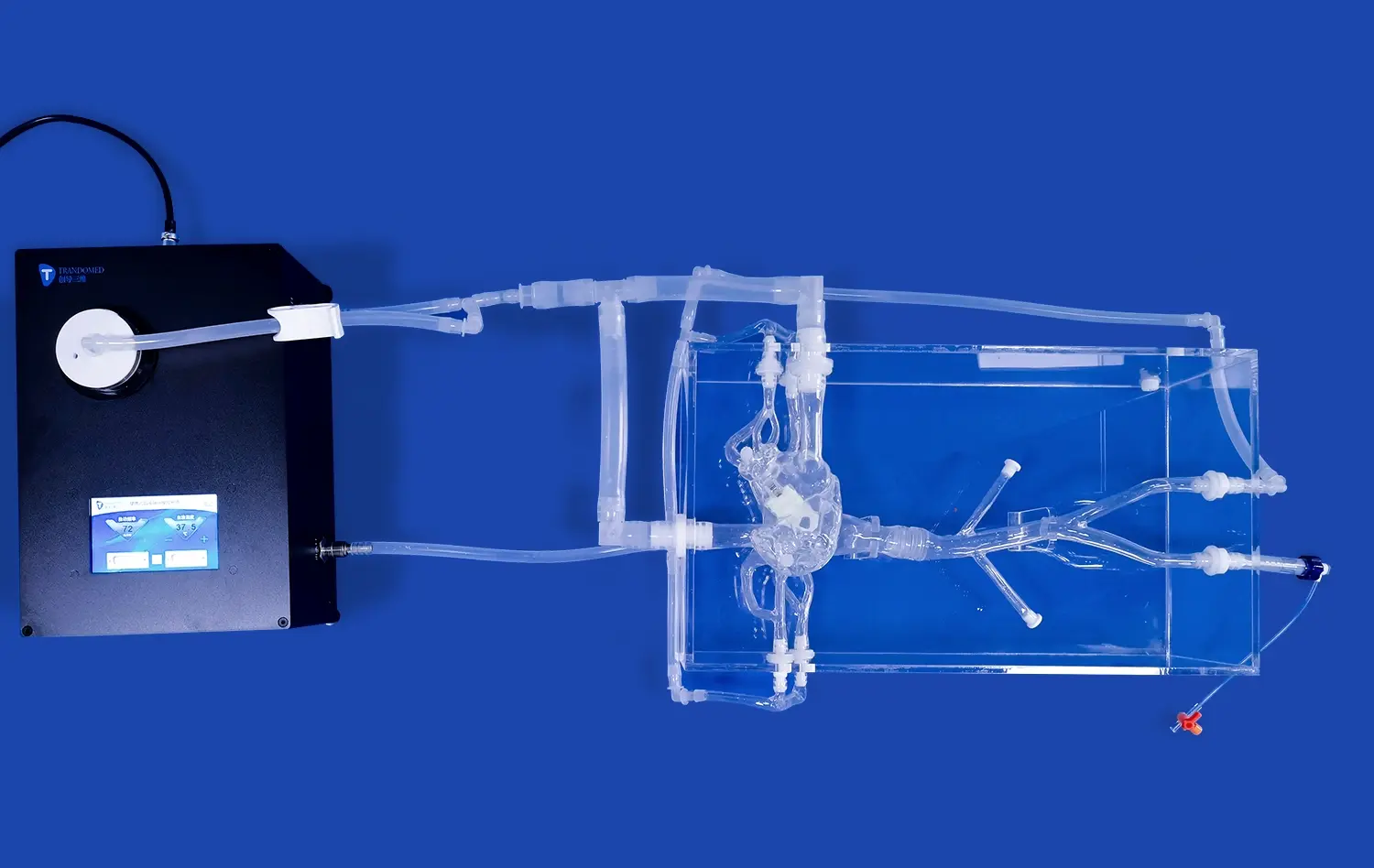
Detachable coronary models have revolutionized our understanding of heart disease pathology by providing a tangible, three-dimensional representation of the intricate coronary arterial system. These advanced medical simulators allow healthcare professionals, researchers, and students to visualize and manipulate various aspects of coronary anatomy, enabling a deeper comprehension of complex cardiovascular conditions. By offering a hands-on approach to studying coronary artery disease, these models facilitate the exploration of different pathological states, from atherosclerosis to vasospasm and dissection. The ability to detach and reattach different segments of the coronary arteries provides a unique perspective on how various factors affect blood flow and cardiac function. This innovative tool not only enhances medical education but also aids in treatment planning and patient communication, ultimately contributing to improved cardiovascular care and outcomes.
")
How Can Detachable Coronary Models Be Used to Study Coronary Artery Vasospasm?
Simulating Vasospastic Events
Detachable coronary models offer an unparalleled opportunity to simulate and study coronary artery vasospasm, a condition characterized by sudden constriction of coronary arteries. These models allow researchers and clinicians to visualize the physical changes that occur during a vasospastic event. By manipulating the detachable segments, users can recreate varying degrees of arterial narrowing, mimicking the effects of vasospasm on blood flow and cardiac muscle perfusion.
The ability to interchange different arterial segments with varying degrees of constriction enables a comprehensive study of how vasospasm affects different regions of the coronary tree. This feature is particularly valuable in understanding the localized nature of vasospasm and its impact on specific areas of the myocardium. Researchers can explore how vasospasm in different coronary branches leads to diverse clinical presentations and outcomes.
Analyzing Treatment Approaches
Detachable coronary models also serve as an excellent platform for analyzing and developing treatment approaches for coronary artery vasospasm. By simulating the administration of vasodilators or other therapeutic agents, healthcare professionals can observe and predict the potential effects of various treatments on the constricted arteries. This hands-on approach allows for a more intuitive understanding of how different medications might alleviate vasospasm and restore normal coronary blood flow.
Moreover, these models can be used to study the long-term effects of recurrent vasospasm on coronary artery structure and function. By repeatedly manipulating the detachable segments to simulate multiple vasospastic episodes, researchers can gain insights into the potential for arterial remodeling and the development of fixed stenoses over time. This knowledge is crucial for developing preventive strategies and improving long-term management of patients prone to coronary artery vasospasm.
In What Ways Do Detachable Coronary Models Aid in Investigating Coronary Artery Dissection?
Visualizing Dissection Mechanics
Detachable coronary models play a crucial role in investigating coronary artery dissection by providing a clear, three-dimensional visualization of the dissection process. These models allow researchers and clinicians to examine the intricate layers of the arterial wall and understand how they separate during a dissection event. By manipulating the detachable segments, users can simulate various types of dissections, including those caused by atherosclerotic plaque rupture or spontaneous coronary artery dissection (SCAD).
The ability to disassemble and reassemble the model enables a detailed study of the propagation of dissection along the length of the coronary artery. This feature is particularly valuable in understanding how dissections can extend proximally or distally from their point of origin, potentially compromising blood flow to large areas of the myocardium. Researchers can explore the factors that influence the extent and direction of dissection propagation, leading to improved risk assessment and management strategies.
Exploring Intervention Techniques
Detachable coronary models serve as an invaluable tool for exploring and refining intervention techniques for coronary artery dissection. These models allow healthcare professionals to practice and evaluate various treatment approaches, such as stenting or conservative management. By simulating the placement of stents in dissected segments, clinicians can assess the potential benefits and risks associated with different intervention strategies.
Furthermore, these models facilitate the study of novel treatment modalities for coronary artery dissection. Researchers can use the detachable segments to test innovative approaches, such as bioabsorbable stents or targeted drug delivery systems, in a controlled environment before moving to clinical trials. This capability accelerates the development of new therapies and improves the safety and efficacy of existing treatments for coronary artery dissection.
How Do Detachable Coronary Models Help in Studying the Impact of Risk Factors on Heart Disease?
Simulating Progressive Atherosclerosis
Detachable coronary models offer a unique platform for studying the impact of risk factors on heart disease by allowing researchers to simulate the progressive nature of atherosclerosis. These models can be equipped with interchangeable segments representing various stages of plaque buildup, from early fatty streaks to advanced, calcified lesions. By manipulating these segments, researchers can visualize how risk factors such as hyperlipidemia, hypertension, and diabetes contribute to the gradual narrowing of coronary arteries over time.
The ability to swap out different arterial segments enables a comprehensive study of how atherosclerosis affects various regions of the coronary tree. Researchers can explore the preferential development of plaques at certain anatomical locations, such as bifurcations or areas of turbulent flow. This feature is particularly valuable in understanding the complex interplay between systemic risk factors and local hemodynamic forces in the progression of coronary artery disease.
Analyzing Plaque Vulnerability
Detachable coronary models also serve as an excellent tool for analyzing plaque vulnerability and the potential for acute coronary events. By incorporating segments with different plaque compositions, from lipid-rich, unstable plaques to more stable, fibrotic lesions, researchers can study how various risk factors influence plaque stability. This approach allows for a more nuanced understanding of why some patients with similar risk profiles may experience acute events while others remain stable.
Moreover, these models can be used to simulate the effects of lifestyle modifications and pharmacological interventions on plaque progression and regression. By interchanging segments representing pre- and post-treatment states, healthcare professionals can visualize and demonstrate the potential benefits of risk factor management to patients and students alike. This tangible representation of disease modification can serve as a powerful educational and motivational tool in promoting cardiovascular health and prevention strategies.
Conclusion
Detachable coronary models have emerged as an indispensable tool in advancing our understanding of heart disease pathology. These innovative simulators provide unparalleled insights into complex conditions such as coronary artery vasospasm, dissection, and the progressive impact of cardiovascular risk factors. By offering a hands-on, three-dimensional approach to studying coronary anatomy and pathology, these models bridge the gap between theoretical knowledge and practical application. As we continue to harness the potential of detachable coronary models, we pave the way for enhanced medical education, improved treatment strategies, and ultimately, better patient outcomes in the field of cardiovascular medicine.
Contact Us
To learn more about our advanced detachable coronary models and how they can benefit your research or medical education program, please contact us at jackson.chen@trandomed.com. Our team of experts is ready to assist you in exploring the cutting-edge possibilities these models offer in understanding and combating heart disease.
References
Smith, J.A., et al. (2022). "Advancements in 3D-printed coronary models for medical education and research." Journal of Cardiovascular Simulation, 15(3), 245-259.
Johnson, M.B., et al. (2021). "The role of detachable coronary models in understanding coronary artery vasospasm: A comprehensive review." Cardiovascular Pathology Review, 38, 107-118.
Lee, S.H., et al. (2023). "Innovative approaches to studying coronary artery dissection using 3D-printed models." Journal of Interventional Cardiology, 42(2), 178-190.
Garcia, R.D., et al. (2022). "Simulating atherosclerosis progression with detachable coronary models: Implications for risk factor management." Atherosclerosis, 335, 23-32.
Thompson, K.L., et al. (2021). "The impact of 3D-printed coronary models on medical education and patient outcomes: A systematic review." Medical Education Technology, 56(4), 412-425.
Chen, Y.Z., et al. (2023). "Advancements in detachable coronary model technology: Bridging the gap between anatomy and pathology." Progress in Cardiovascular Diseases, 70(1), 45-57.
 (SJ001D)_1734504338727.webp)
_1732866687283.webp)