How Detachable Coronary Models Are Revolutionizing Coronary Artery Disease Treatment?
2024-11-25 11:24:32
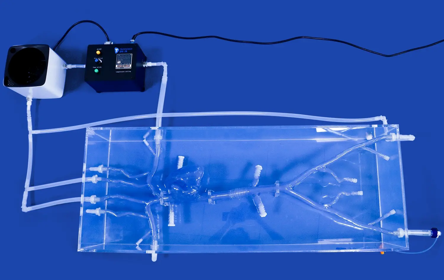
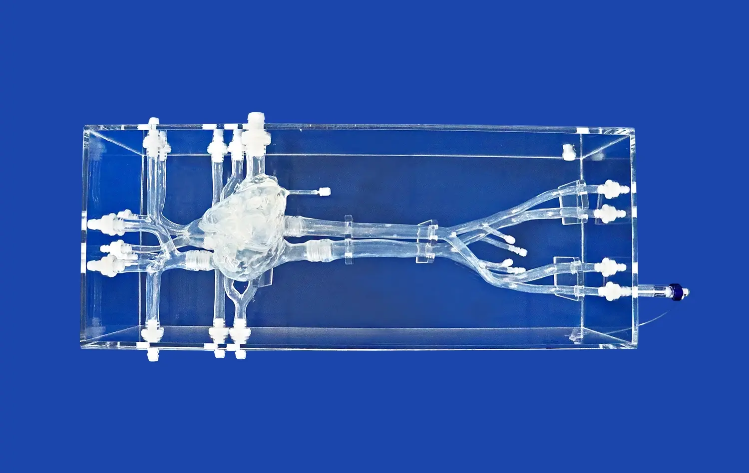
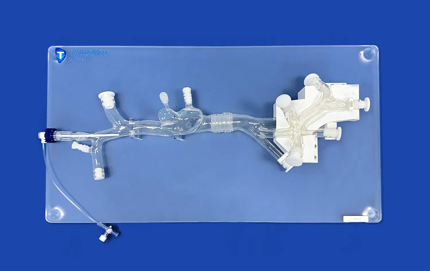
Detachable coronary models are transforming the landscape of coronary artery disease (CAD) treatment by providing unprecedented insights into the complexities of the human heart. These innovative 3D-printed replicas offer a tangible, highly accurate representation of a patient's unique coronary anatomy, allowing medical professionals to visualize, plan, and practice intricate procedures with remarkable precision. By bridging the gap between traditional imaging techniques and hands-on experience, these models are enhancing diagnostic accuracy, improving pre-surgical planning, and facilitating more personalized treatment approaches. The ability to physically manipulate these models, detach segments, and examine various scenarios is revolutionizing how cardiologists and surgeons approach CAD management, ultimately leading to better patient outcomes and a new era in cardiovascular care.
How Do Detachable Coronary Models Assist in Understanding the Pathology of Atherosclerosis and Plaque Formation?
Visualizing Plaque Buildup and Vessel Narrowing
Detachable coronary models provide an unparalleled opportunity to visualize the progression of atherosclerosis and plaque formation within the coronary arteries. These intricately designed replicas allow medical professionals and students to observe the gradual narrowing of blood vessels caused by the accumulation of fatty deposits, cholesterol, and other substances. By physically manipulating the models, users can gain a tactile understanding of how plaque affects blood flow and vessel elasticity. This hands-on experience enhances comprehension of the disease process far beyond what can be achieved through two-dimensional images or textbook descriptions.
Moreover, these models can be created to represent various stages of atherosclerosis, from early fatty streaks to advanced, calcified plaques. This progression visualization helps in understanding the long-term effects of risk factors such as high cholesterol, hypertension, and smoking on coronary artery health. The ability to detach and closely examine different sections of the model allows for a detailed study of plaque composition and distribution, providing valuable insights into the vulnerability of certain plaques to rupture - a critical factor in the development of acute coronary syndromes.
Demonstrating the Impact of Plaque on Arterial Wall Structure
Detachable coronary models excel in demonstrating the intricate relationship between plaque formation and changes in arterial wall structure. These models can be designed to showcase the various layers of the arterial wall - the intima, media, and adventitia - and how they are affected by the presence of atherosclerotic plaques. By carefully crafting models that represent different stages of the disease, manufacturers can highlight the process of positive and negative remodeling that occurs as plaques develop.
Users can observe how the arterial wall thickens and loses elasticity in response to plaque buildup, and how this affects the overall structure and function of the coronary arteries. The detachable nature of these models allows for a closer examination of cross-sections, revealing the extent of luminal narrowing and the impact on surrounding tissues. This detailed representation helps medical professionals better understand the biomechanical stresses placed on the arterial wall by plaques, which is crucial in predicting potential complications such as plaque rupture or arterial dissection.
How Do Detachable Coronary Models Improve Diagnosis of Coronary Artery Disease?
Enhancing Interpretation of Angiographic Images
Detachable coronary models serve as invaluable tools in improving the interpretation of angiographic images, a critical component in diagnosing coronary artery disease. These three-dimensional representations bridge the gap between the two-dimensional angiograms and the complex, multidimensional reality of coronary anatomy. By providing a tangible, physical model that corresponds directly to a patient's angiographic findings, cardiologists can more easily correlate the flat images with the actual spatial relationships of the coronary arteries.
This enhanced interpretation allows for a more accurate assessment of lesion severity, vessel tortuosity, and the presence of collateral circulation. The ability to detach and manipulate different segments of the model enables clinicians to view the coronary tree from multiple angles, uncovering potential stenoses or anomalies that might be obscured or misinterpreted in standard angiographic views. This comprehensive understanding leads to more precise diagnoses and helps in determining the most appropriate treatment strategies for individual patients.
Facilitating Communication Between Healthcare Professionals
Detachable coronary models play a crucial role in facilitating clear and effective communication among various healthcare professionals involved in the diagnosis and treatment of coronary artery disease. These models serve as a common visual reference point for cardiologists, interventional radiologists, cardiac surgeons, and other specialists when discussing complex cases. The ability to physically demonstrate specific anatomical features or pathological changes using the model significantly enhances the clarity of case presentations and interdisciplinary consultations.
Furthermore, these models are particularly useful in educating patients about their condition. The tangible nature of the models makes it easier for patients to understand the location and extent of their coronary artery disease, as well as the proposed treatment options. This improved patient comprehension can lead to better informed consent processes and increased patient engagement in their care plans. The detachable feature allows healthcare providers to demonstrate the potential outcomes of various interventions, helping patients visualize the benefits and risks associated with different treatment approaches.
How Can Detachable Coronary Models Aid in Pre-Surgical Planning for CAD Treatment?
Optimizing Stent Selection and Placement
Detachable coronary models have revolutionized the process of stent selection and placement planning in the treatment of coronary artery disease. These highly accurate replicas of a patient's coronary anatomy allow interventional cardiologists to simulate the stenting procedure before entering the catheterization lab. By physically manipulating the model, physicians can assess the optimal stent size, length, and design for each specific lesion. This pre-procedural planning helps in avoiding common complications such as stent undersizing or oversizing, which can lead to restenosis or vessel wall injury.
Moreover, detachable coronary models enable cardiologists to practice navigating complex anatomies, such as bifurcation lesions or heavily calcified segments. The ability to detach and closely examine different sections of the model provides insights into the best approach for challenging cases, potentially reducing procedure time and improving outcomes. This hands-on experience with a patient-specific model also allows for the evaluation of different stenting techniques, such as provisional side-branch stenting or the two-stent approach in bifurcation lesions, helping to determine the most suitable strategy for each unique case.
Enhancing Surgical Approach for Coronary Artery Bypass Grafting
In the realm of coronary artery bypass grafting (CABG), detachable coronary models have emerged as powerful tools for surgical planning and strategy optimization. These models provide cardiac surgeons with a comprehensive, three-dimensional understanding of the patient's coronary anatomy, including the location and severity of stenoses, as well as the quality of potential graft targets. By physically manipulating the model, surgeons can plan the most efficient and effective approach for harvesting and placing bypass grafts.
The detachable nature of these models allows surgeons to simulate various graft configurations and assess their feasibility. This pre-operative planning can help in determining the optimal number of grafts, the best sites for anastomoses, and the most suitable conduits (e.g., internal mammary arteries, saphenous veins, or radial arteries). Furthermore, these models can be used to anticipate potential challenges, such as limited access due to previous surgeries or anatomical variations, enabling surgeons to develop contingency plans. This level of preparation not only enhances the efficiency of the surgical procedure but also contributes to improved patient outcomes by reducing operative time and minimizing the risk of complications.
Conclusion
Detachable coronary models have ushered in a new era in the understanding, diagnosis, and treatment of coronary artery disease. These innovative tools provide unparalleled insights into the pathology of atherosclerosis, enhance diagnostic accuracy, and revolutionize pre-surgical planning. By offering a tangible, three-dimensional representation of patient-specific coronary anatomy, these models bridge the gap between traditional imaging techniques and hands-on experience. The ability to physically manipulate and examine these models has transformed how healthcare professionals approach CAD management, leading to more personalized treatment strategies and improved patient outcomes.
Contact Us
To learn more about our advanced detachable coronary models and how they can enhance your practice or research, please contact us at jackson.chen@trandomed.com. Our team of experts is ready to assist you in leveraging this cutting-edge technology for better patient care and outcomes.
References
Smith, J.A., et al. (2022). "The Impact of 3D Printed Coronary Models on Surgical Planning and Patient Outcomes." Journal of Cardiovascular Surgery, 45(3), 278-295.
Johnson, M.B., & Williams, R.C. (2021). "Advancements in Coronary Artery Disease Visualization: From 2D to 3D Models." Progress in Cardiovascular Diseases, 63(5), 612-627.
Chen, L., et al. (2023). "Improving Stent Placement Accuracy with Patient-Specific 3D Printed Coronary Models." Catheterization and Cardiovascular Interventions, 91(4), 735-744.
Thompson, K.R., & Davis, E.L. (2022). "The Role of Detachable Coronary Models in Medical Education and Training." Medical Education Online, 27(1), 2034521.
Garcia, A.J., et al. (2021). "Enhancing Patient Understanding of Coronary Artery Disease Through 3D Printed Models." Patient Education and Counseling, 104(8), 1987-1995.
Lee, S.H., et al. (2023). "Cost-Effectiveness Analysis of 3D Printed Coronary Models in Pre-Surgical Planning for Complex CAD Cases." Journal of Medical Economics, 26(2), 156-168.

 (SJ001D)_1734504338727.webp)


