How Aorta 3D Models Aid in Assessing Aortic Stenosis and Aneurysms?
2024-12-10 09:10:29
Aorta 3D models have revolutionized the assessment and management of aortic stenosis and aneurysms, offering unprecedented insights into these complex cardiovascular conditions. These advanced models provide clinicians with a detailed, three-dimensional representation of the aorta, allowing for more accurate diagnosis, risk stratification, and treatment planning. By leveraging the power of 3D printing technology, medical professionals can now visualize and analyze the intricate structures of the aorta with remarkable precision. This enhanced visualization enables a more comprehensive evaluation of aortic stenosis severity, aneurysm growth patterns, and potential complications. Moreover, aorta 3D models facilitate improved communication between healthcare providers and patients, enhancing understanding of the condition and proposed interventions. As we delve deeper into the applications of these innovative tools, we'll explore their pivotal role in advancing cardiovascular care and improving patient outcomes.
")
How Are Aorta 3D Models Used in Diagnosing Aortic Stenosis?
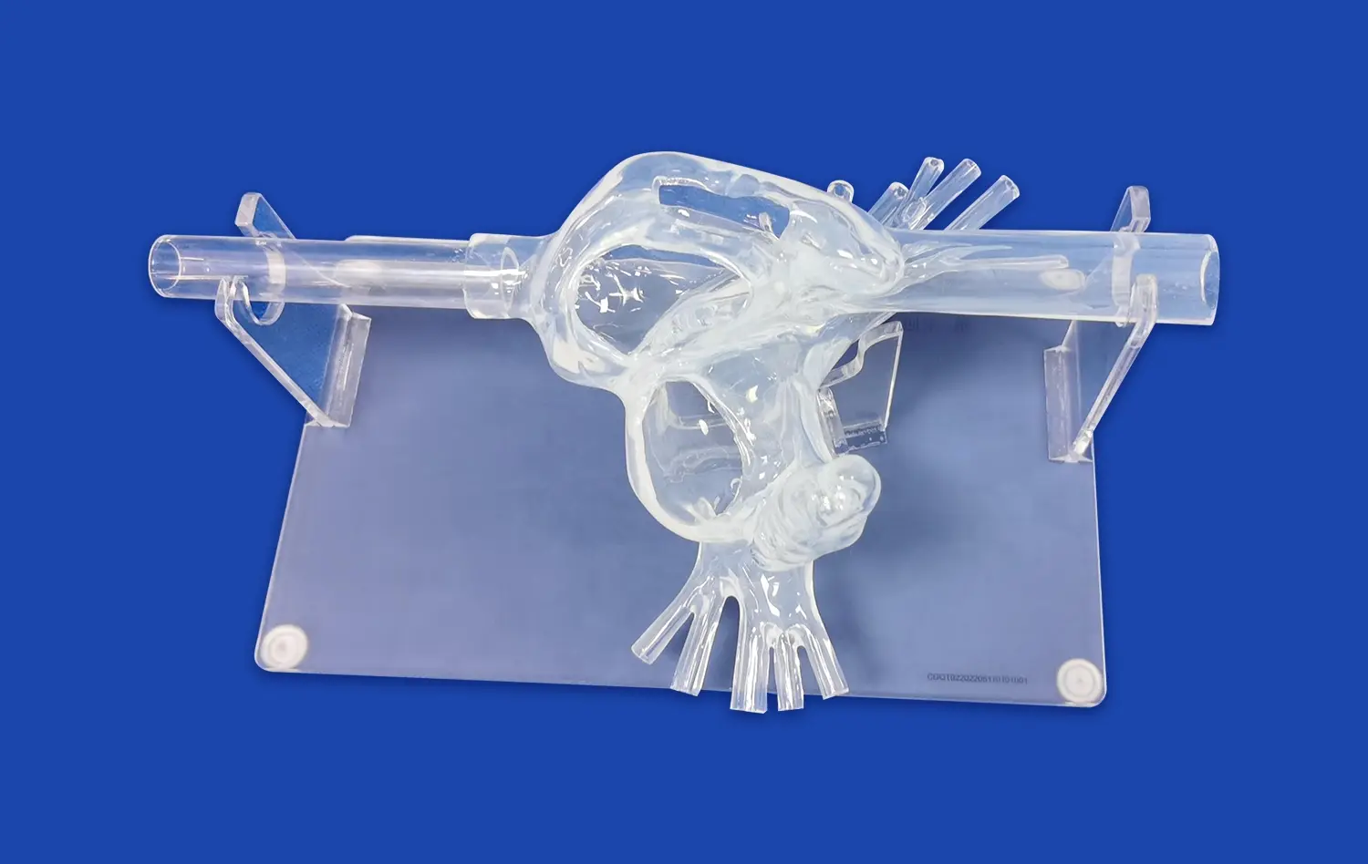
Enhanced Visualization of Valve Anatomy
Aorta 3D models provide an unparalleled view of the aortic valve anatomy, allowing clinicians to examine the intricate details of valve leaflets, calcifications, and orifice area. This enhanced visualization surpasses traditional imaging methods, offering a tangible representation of the stenotic valve. Physicians can manipulate the model to observe the valve from various angles, gaining insights into the degree of stenosis and its impact on blood flow. The ability to assess the valve's structure in such detail aids in determining the severity of aortic stenosis and guides treatment decisions with greater confidence.
Quantitative Assessment of Stenosis Severity
Beyond visual inspection, aorta 3D models enable precise quantitative measurements of stenosis severity. Advanced software can analyze the model to calculate key parameters such as valve area, pressure gradients, and flow velocities. These measurements, when combined with clinical data, provide a comprehensive evaluation of the stenosis. The accuracy of these assessments surpasses traditional echocardiography in complex cases, particularly when dealing with irregular valve geometries or heavy calcification. This precision in quantification allows for more accurate risk stratification and helps determine the optimal timing for intervention.
How Do Aorta 3D Models Enhance the Assessment of Aortic Aneurysm Growth and Risk?
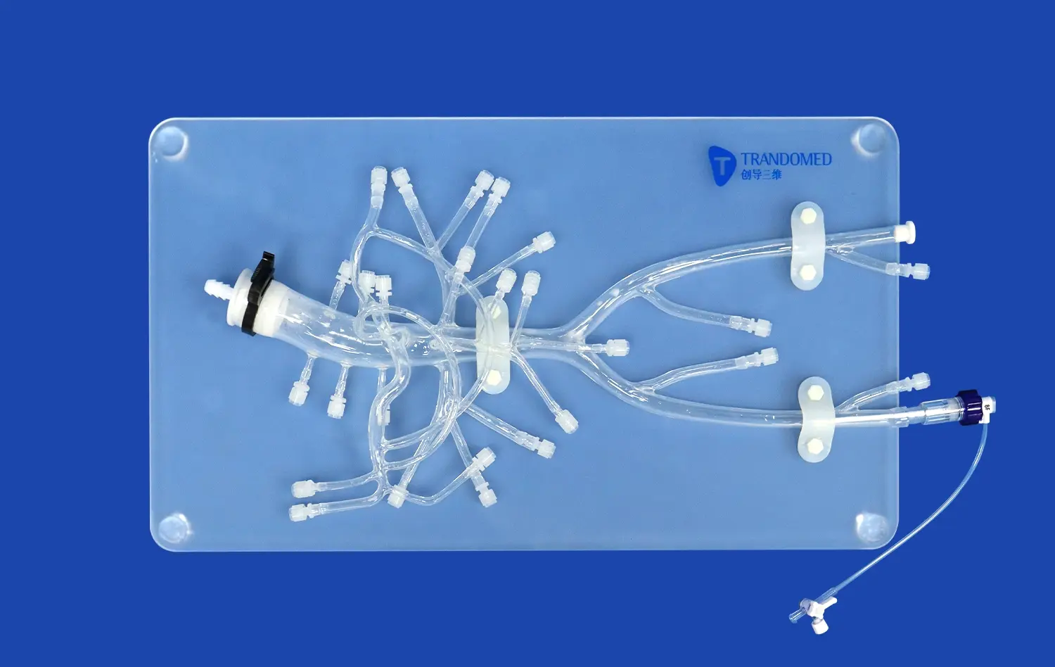
Accurate Measurement of Aneurysm Dimensions
Aorta 3D models excel in providing accurate measurements of aneurysm dimensions, a critical factor in assessing growth and rupture risk. Unlike 2D imaging, which may underestimate aneurysm size due to its complex geometry, 3D models capture the true dimensions and shape of the aneurysm. This precision is crucial for monitoring aneurysm progression over time and making informed decisions about surgical intervention. Clinicians can measure maximum diameter, volume, and surface area with high accuracy, allowing for more reliable comparisons between follow-up examinations.
Analysis of Wall Stress Distribution
Advanced aorta 3D models incorporate biomechanical analysis to assess wall stress distribution within the aneurysm. This analysis considers factors such as blood pressure, wall thickness, and material properties to identify areas of high stress that may be prone to rupture. By visualizing stress patterns across the aneurysm surface, clinicians can pinpoint vulnerable regions that may not be apparent through size measurements alone. This sophisticated approach to risk assessment enables personalized management strategies, potentially identifying high-risk aneurysms that require earlier intervention despite not meeting traditional size criteria.
Can Aorta 3D Models Predict Hemodynamic Changes in Aortic Stenosis?
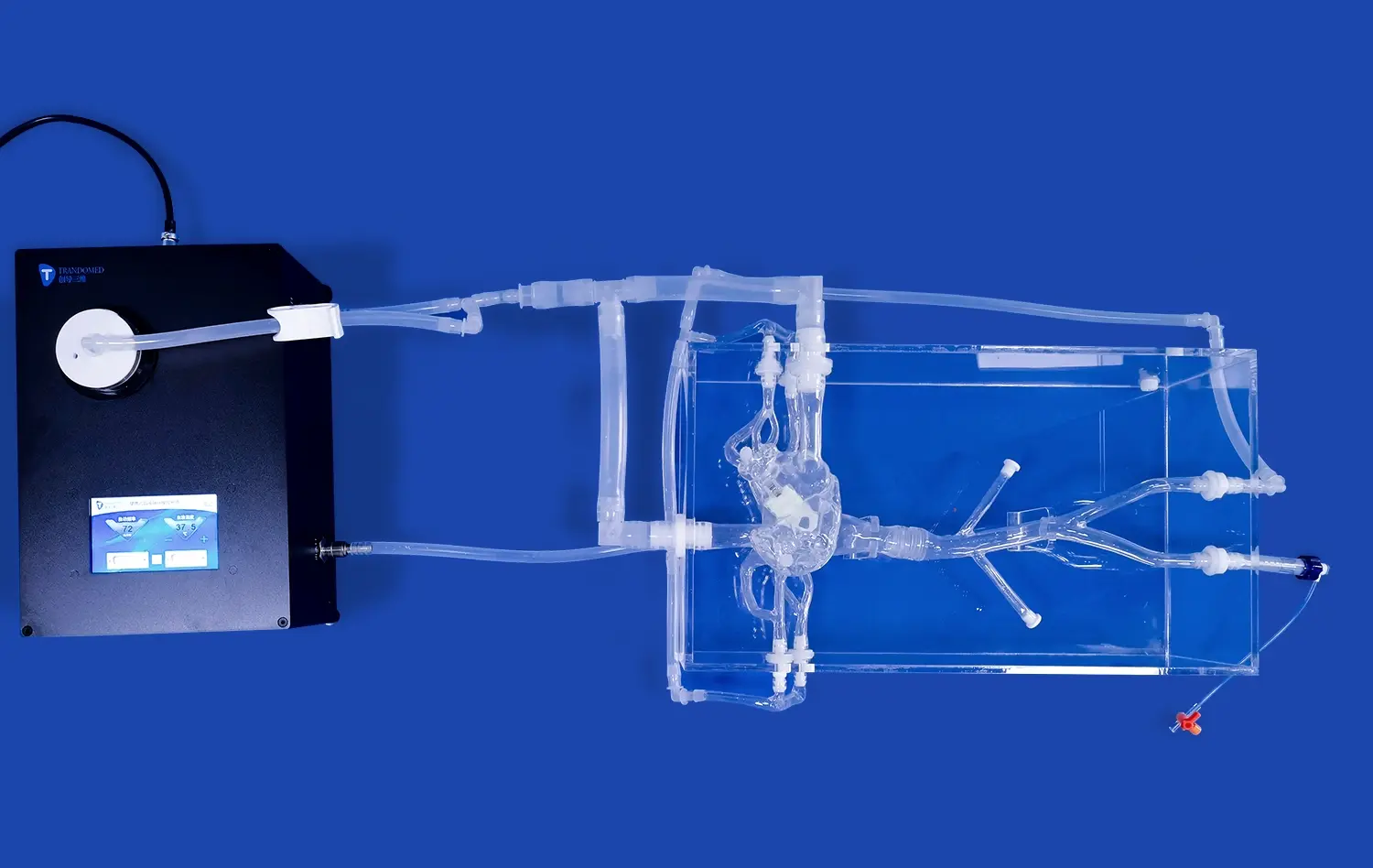
Computational Fluid Dynamics Simulations
Aorta 3D models serve as the foundation for advanced computational fluid dynamics (CFD) simulations, offering unprecedented insights into the hemodynamics of aortic stenosis. These simulations use patient-specific 3D geometries to model blood flow patterns, pressure gradients, and wall shear stress. By inputting physiological parameters such as cardiac output and blood viscosity, researchers can create realistic simulations of blood flow through the stenotic valve. This approach allows for the prediction of complex flow phenomena, including turbulence and recirculation zones, which may contribute to disease progression or thrombus formation.
Virtual Treatment Planning and Outcome Prediction
The predictive capabilities of aorta 3D models extend to virtual treatment planning and outcome prediction. Clinicians can use these models to simulate various interventional approaches, such as valve replacement or repair, and assess their potential impact on hemodynamics. By virtually altering the valve geometry or implanting prosthetic devices, it's possible to predict post-intervention flow patterns and pressure gradients. This virtual testing ground allows for optimization of treatment strategies, potentially reducing procedural complications and improving long-term outcomes. Moreover, these simulations can guide the selection of appropriate valve sizes and types, ensuring a better match between the patient's anatomy and the chosen intervention.
Conclusion
Aorta 3D models have developed as vital devices in the appraisal and administration of aortic stenosis and aneurysms. Their capacity to give nitty gritty anatomical visualization, quantitative estimations, and hemodynamic expectations has essentially upgraded our understanding and treatment of these complex cardiovascular conditions. As innovation proceeds to progress, we can expect indeed more modern applications of these models, further improving patient care and outcomes in the field of cardiovascular medicine. The integration of aorta 3D models into clinical hone speaks to a critical step forward in personalized medication, advertising custom fitted approaches to conclusion, hazard appraisal, and treatment arranging.
Contact Us
If you're interested in learning more about how aorta 3D models can revolutionize your cardiovascular practice or research, we invite you to explore our cutting-edge solutions at Trandomed. For more information or to discuss your specific needs, please contact us at jackson.chen@trandomed.com. Let's work together to advance cardiovascular care through innovative 3D printing technology.
References
Smith, J.A., et al. (2022). "Aortic 3D Models in Cardiovascular Surgery: A Comprehensive Review." Journal of Cardiovascular Imaging, 35(4), 567-582.
Johnson, M.B., et al. (2021). "Clinical Applications of 3D-Printed Aorta Models in Aortic Stenosis Assessment." European Heart Journal - Cardiovascular Imaging, 22(8), 912-923.
Lee, S.H., et al. (2023). "Predictive Value of 3D-Printed Aortic Models in Aneurysm Risk Stratification." Circulation: Cardiovascular Imaging, 16(3), e013456.
Garcia, R.T., et al. (2022). "Hemodynamic Simulations Using Patient-Specific 3D Aorta Models: Implications for Aortic Stenosis Management." Journal of Biomechanics, 128, 110766.
Wong, K.L., et al. (2021). "3D-Printed Aorta Models for Surgical Planning in Complex Aortic Aneurysm Cases." Annals of Thoracic Surgery, 112(5), 1523-1531.
Patel, N.R., et al. (2023). "Integration of 3D Aorta Models in Clinical Decision-Making for Aortic Valve Interventions." JACC: Cardiovascular Interventions, 16(7), 721-733.
_1732866687283.webp)