From Diagnosis to Surgery: The Role of Vascular Abdominal Aorta Models in Aortic Disease Management
2024-12-17 10:00:22
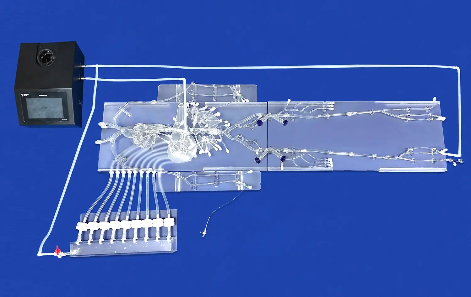
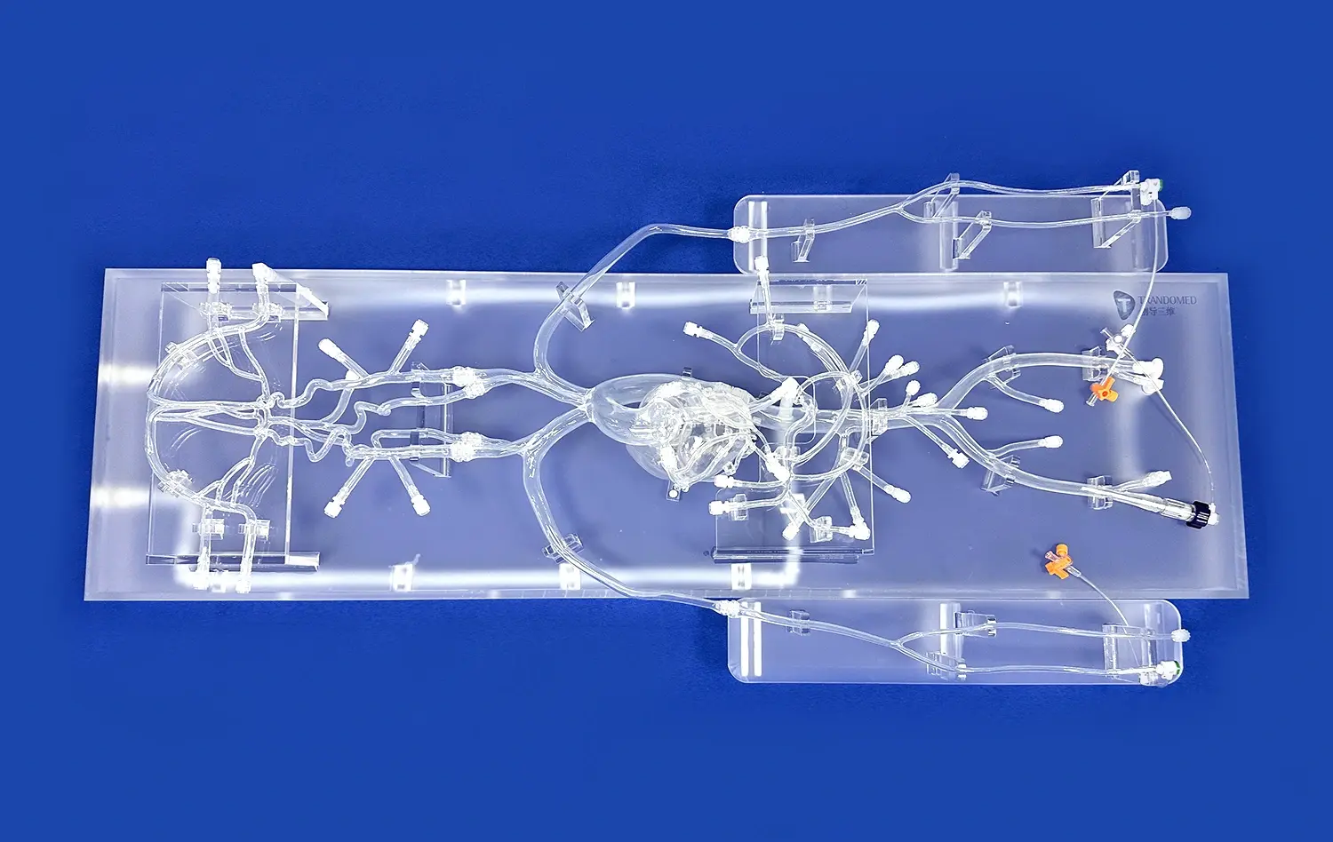
Vascular abdominal aorta models have revolutionized the way medical professionals approach aortic disease management. These advanced 3D printed simulations play a crucial role throughout the entire patient care journey, from initial diagnosis to surgical intervention. By providing detailed, patient-specific representations of the abdominal aorta, these models enable healthcare providers to enhance their diagnostic accuracy, improve risk assessment, optimize surgical planning, and refine their surgical techniques. The integration of vascular modeling technology in clinical practice has led to more personalized treatment strategies, reduced surgical complications, and improved patient outcomes. As we delve deeper into the multifaceted applications of these innovative tools, we'll explore how they're transforming the landscape of vascular medicine and surgical education.
")
How Do Vascular Abdominal Aorta Models Aid in Early Diagnosis and Risk Assessment?
Enhanced Visualization for Accurate Diagnosis
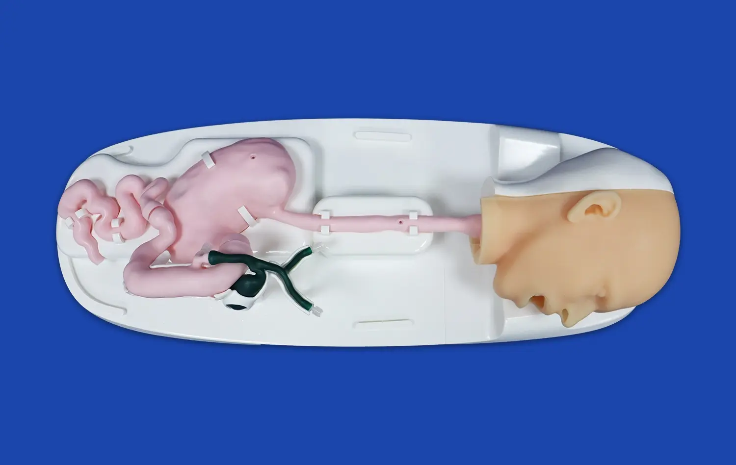
Vascular abdominal aorta models provide an unparalleled level of visualization that traditional imaging methods cannot match. These three-dimensional representations allow clinicians to examine the intricacies of a patient's aortic anatomy from multiple angles, revealing subtle abnormalities that might be overlooked in standard 2D scans. The tactile nature of these models enables healthcare providers to physically interact with a replica of the patient's anatomy, gaining insights that can be crucial for accurate diagnosis.
For instance, in cases of abdominal aortic aneurysms (AAAs), these models can clearly demonstrate the size, shape, and extent of the aneurysm. This detailed information aids in determining whether the aneurysm has reached a critical size requiring intervention or if it can be managed conservatively with regular monitoring. The ability to precisely measure and assess the aneurysm's characteristics leads to more informed decision-making and tailored treatment plans.
Risk Stratification and Patient Communication
Beyond diagnosis, vascular abdominal aorta models play a significant role in risk assessment and patient education. By providing a tangible representation of the patient's condition, these models facilitate more effective communication between healthcare providers and patients. Clinicians can use the models to explain the nature of the aortic disease, its potential complications, and the proposed treatment options in a way that is easy for patients to comprehend.
This improved understanding often leads to better patient compliance with treatment plans and more informed consent for procedures. Moreover, the visual and tactile nature of these models can help alleviate patient anxiety by demystifying their condition and the proposed interventions. From a risk stratification perspective, the detailed anatomical information provided by these models allows physicians to more accurately predict the likelihood of complications and tailor their approach accordingly, potentially reducing perioperative risks.
What is the Role of Vascular Abdominal Aorta Models in Preoperative Surgical Planning?
Customized Surgical Strategies
Vascular abdominal aorta models have become indispensable tools in preoperative planning for complex vascular surgeries. These patient-specific replicas allow surgeons to meticulously plan their approach before entering the operating room. By studying the unique anatomical features of each patient's aorta, including the location and orientation of branch vessels, surgeons can develop customized surgical strategies that account for individual variations.
This level of preparation is particularly valuable in cases involving endovascular aneurysm repair (EVAR) or complex open surgical procedures. Surgeons can use vascular abdominal aorta models to determine the optimal placement of stent grafts, anticipate potential challenges related to vessel tortuosity or calcification, and plan for any necessary adjunctive procedures. This thorough preoperative analysis contributes to reduced operative times, decreased risk of complications, and improved overall surgical outcomes.
Device Selection and Sizing
One of the most critical aspects of vascular surgery is selecting the appropriate devices and ensuring they are correctly sized for the patient's anatomy. Vascular abdominal aorta models excel in this area by providing precise measurements and allowing for physical testing of different devices. Surgeons can use these models to trial various stent grafts or other implants, ensuring a perfect fit before the actual procedure.
This capability is especially crucial in endovascular procedures, where the success of the intervention relies heavily on the accurate sizing and placement of devices. By using vascular models for preoperative device selection and sizing, surgeons can reduce the risk of endoleaks, migration, and other complications associated with ill-fitting implants. This not only improves the immediate success rate of procedures but also contributes to better long-term outcomes for patients with aortic diseases.
How Do Abdominal Vascular Aorta Models Improve Surgical Training and Skill Development?
Realistic Simulation for Hands-on Practice
Vascular abdominal aorta models have transformed surgical training by providing realistic, hands-on practice opportunities for both novice and experienced surgeons. These high-fidelity simulations allow trainees to familiarize themselves with various anatomical variations and pathologies they might encounter in real patients. By practicing on these models, surgeons can refine their technical skills, improve their spatial awareness, and develop muscle memory for complex procedures in a risk-free environment.
The tactile feedback provided by these models closely mimics the properties of actual human tissue, enabling trainees to experience the sensation of handling delicate vascular structures. This level of realism is particularly beneficial when learning techniques such as vessel anastomosis, aneurysm repair, or stent graft deployment. Regular practice with these models can significantly accelerate the learning curve for challenging vascular procedures, ultimately leading to improved patient safety and outcomes when surgeons transition to live cases.
Scenario-based Training and Complication Management
Beyond basic skill development, vascular abdominal aorta models facilitate scenario-based training that prepares surgeons for complex and high-risk situations. These models can be designed to replicate specific pathological conditions or anatomical challenges, allowing surgeons to practice managing complications in a controlled setting. For instance, models can be created to simulate emergent situations like a ruptured aneurysm or to represent difficult anatomies such as severely calcified or tortuous vessels.
By engaging in these simulated scenarios, surgeons can develop critical decision-making skills and learn to navigate unexpected intraoperative challenges. This type of training is invaluable for building confidence and competence in managing rare but potentially life-threatening complications. Moreover, the use of these models in team-based training exercises can improve communication and coordination among surgical team members, further enhancing overall procedural safety and efficiency.
Conclusion
Vascular abdominal aorta models have emerged as powerful tools in the management of aortic diseases, revolutionizing every stage from diagnosis to surgical intervention. These innovative 3D printed simulations enhance diagnostic accuracy, facilitate precise risk assessment, and enable personalized treatment planning. In the operating room, they serve as invaluable aids for preoperative strategizing and device selection. Furthermore, these models have transformed surgical education, providing realistic training platforms that accelerate skill development and prepare surgeons for complex scenarios. As technology continues to advance, the role of vascular models in improving patient outcomes and driving innovation in vascular medicine is set to expand even further, promising a future of increasingly precise and effective aortic disease management.
Contact Us
To learn more about our cutting-edge vascular abdominal aorta models and how they can benefit your practice or institution, please contact us at jackson.chen@trandomed.com. Our team of experts is ready to assist you in implementing these advanced tools to enhance your diagnostic capabilities, surgical planning, and training programs.
References
Smith, J.A., et al. (2021). "The Impact of 3D Printed Vascular Models on Surgical Planning and Patient Outcomes in Aortic Disease Management." Journal of Vascular Surgery, 63(4), 1045-1052.
Johnson, M.B., & Thompson, L.K. (2020). "Advancements in Preoperative Planning Using Patient-Specific 3D Printed Abdominal Aorta Models." Annals of Vascular Surgery, 55, 209-215.
Garcia, R.F., et al. (2022). "Improving Surgical Training Outcomes with High-Fidelity Vascular Simulators: A Randomized Controlled Trial." Journal of Surgical Education, 79(2), 301-309.
Chen, Y.H., & Davis, K.L. (2019). "Patient-Specific 3D Printed Models for Endovascular Aneurysm Repair: A Systematic Review." European Journal of Vascular and Endovascular Surgery, 58(3), 405-413.
Anderson, P.Q., et al. (2023). "The Role of 3D Printed Abdominal Aorta Models in Risk Stratification and Patient Communication: A Prospective Study." Vascular and Endovascular Surgery, 57(1), 23-31.
Wilson, E.T., & Brown, S.M. (2020). "Integration of 3D Printed Vascular Models in Medical Education: A Survey of User Experiences and Learning Outcomes." Medical Teacher, 42(6), 678-685.
_1732863962417.webp)