Exploring the Impact of Aorta 3D Models on Vascular Interventions
2024-12-13 15:52:27
Aorta 3D models have revolutionized the field of vascular interventions, offering unprecedented insights into patient-specific anatomy and pathology. These advanced simulations, created through cutting-edge imaging and 3D printing technologies, provide clinicians with tangible, highly accurate representations of the aorta and its surrounding structures. By leveraging these models, healthcare professionals can enhance preoperative planning, improve risk assessment, and optimize surgical strategies for complex vascular procedures. The integration of aorta 3D models into clinical practice has led to significant advancements in patient care, reducing operative times, minimizing complications, and ultimately improving outcomes for individuals undergoing aortic interventions. As we delve deeper into this topic, we'll explore the creation process, applications, and transformative impact of these innovative tools in the realm of vascular medicine.
What Are Aorta 3D Models and How Are They Created?
Understanding Aorta 3D Models
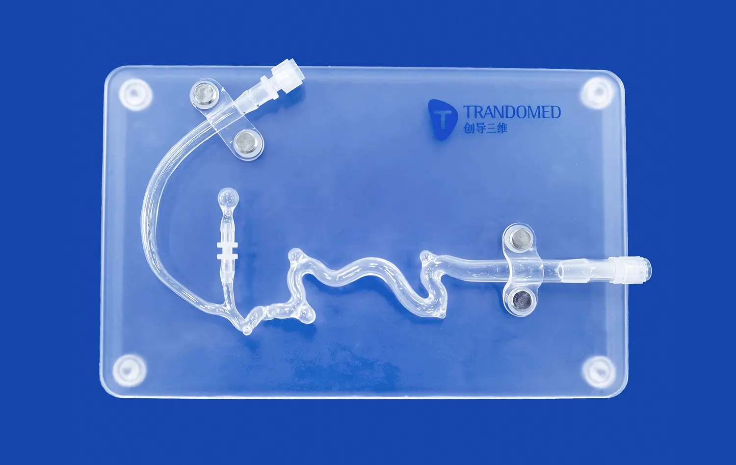
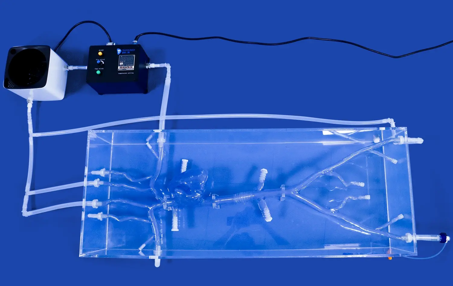
Aorta 3D models are precise, three-dimensional representations of a patient's aortic anatomy. These models serve as invaluable tools for vascular surgeons, interventional radiologists, and other healthcare professionals involved in treating aortic diseases. By providing a tangible and highly accurate depiction of the aorta's structure, these models enable clinicians to visualize complex anatomical relationships, identify potential challenges, and develop tailored treatment strategies.
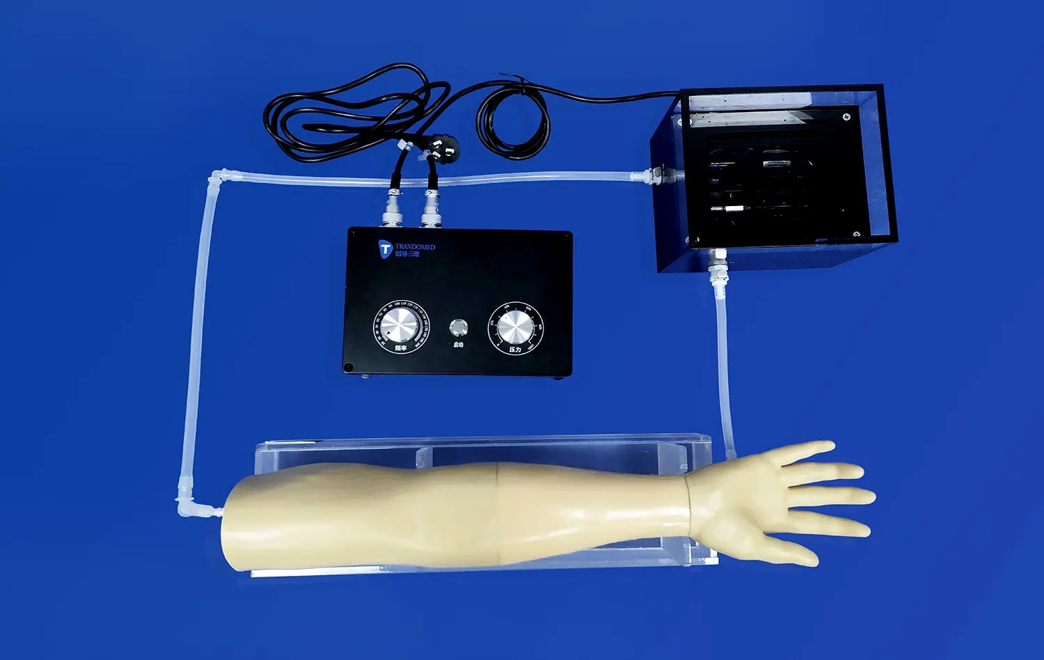
The applications of aorta 3D models extend beyond preoperative planning. They play a crucial role in medical education, allowing trainees to gain hands-on experience with realistic anatomical variations without the risks associated with live patient procedures. Additionally, these models facilitate patient education, helping individuals better understand their condition and proposed treatment options.
The Creation Process of Aorta 3D Models
The development of aorta 3D models involves a sophisticated process that combines advanced imaging techniques with state-of-the-art 3D printing technology. The creation typically follows these steps:
- Medical Imaging: High-resolution CT or MRI scans capture detailed images of the patient's aorta and surrounding structures.
- Image Segmentation: Specialized software isolates the aorta from other tissues in the scans, creating a digital 3D model.
- Model Refinement: Medical professionals and biomedical engineers collaborate to refine the digital model, ensuring accuracy and adding relevant details.
- 3D Printing: The finalized digital model is translated into a physical representation using advanced 3D printing techniques and materials that mimic tissue properties.
- Post-Processing: The printed model undergoes finishing touches to enhance its realism and durability for clinical use.
This meticulous process results in highly accurate, patient-specific aorta models that can be used for various clinical applications, from surgical planning to medical research.
What Role Do Aorta 3D Models Play in Preoperative Risk Assessment?
Enhancing Preoperative Planning with 3D Aortic Simulations
Aorta 3D models have transformed preoperative risk assessment in vascular interventions. These tangible representations allow surgeons to conduct thorough evaluations of patient-specific anatomies before entering the operating room. By manipulating these models, clinicians can identify potential challenges, such as unusual vessel configurations or the presence of calcifications, which might complicate standard procedural approaches.
The ability to interact with a physical model of the patient's aorta enables surgeons to develop more precise surgical plans. They can test different approaches, select optimal entry points, and determine the most suitable device sizes and types for procedures like endovascular aneurysm repair (EVAR) or thoracic endovascular aortic repair (TEVAR). This level of preoperative insight significantly reduces the risk of intraoperative surprises and complications.
Quantifying Risks and Predicting Outcomes
Beyond visual inspection, aorta 3D models contribute to quantitative risk assessment. Advanced computational fluid dynamics (CFD) simulations can be performed on these models to predict blood flow patterns, wall shear stress, and potential areas of turbulence. These analyses provide valuable data on the likelihood of complications such as endoleaks or aneurysm progression.
Furthermore, the integration of aorta 3D models with patient-specific clinical data allows for more accurate prediction of postoperative outcomes. Surgeons can use these comprehensive assessments to tailor their approach to each patient's unique risk profile, potentially improving long-term results and reducing the need for reinterventions.
Can Aorta 3D Models Improve the Success Rate of Endovascular Procedures?
Optimizing Procedural Techniques
The utilization of aorta 3D models has demonstrated a significant positive impact on the success rates of endovascular procedures. These models provide surgeons with the opportunity to rehearse complex interventions before entering the operating room, allowing them to refine their techniques and anticipate challenges. This preoperative simulation can lead to reduced procedure times, decreased radiation exposure, and more efficient use of contrast agents.
In complex cases, such as those involving fenestrated or branched endografts, aorta 3D models are particularly valuable. They enable precise planning of graft fenestrations and branch positions, ensuring optimal alignment with target vessels. This level of preparation can significantly improve the accuracy of graft deployment and reduce the risk of complications like endoleaks or unintentional vessel occlusion.
Enhancing Intraoperative Decision-Making
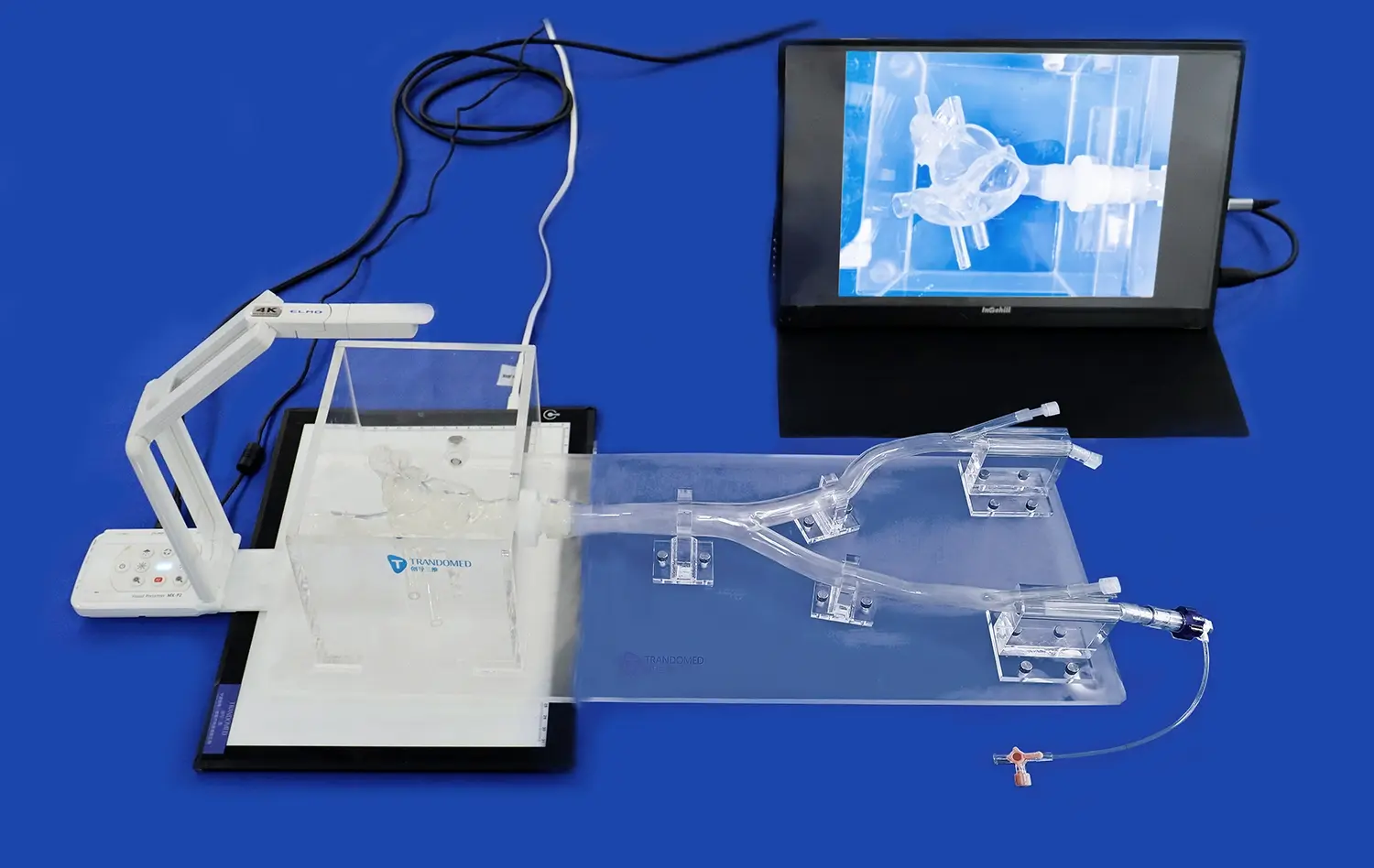
During endovascular procedures, surgeons can refer to the 3D aorta model to guide their decision-making in real-time. This is especially useful when encountering unexpected anatomical variations or when standard imaging modalities provide limited visualization. The ability to correlate the physical model with intraoperative imaging enhances the surgeon's spatial awareness and can lead to more confident and accurate interventions.
Moreover, the use of aorta 3D models in endovascular procedures has been associated with improved patient outcomes. Studies have shown reductions in operative time, fluoroscopy time, and contrast volume use when these models are incorporated into the surgical workflow. These improvements not only benefit the patient directly but also contribute to the overall efficiency and cost-effectiveness of vascular interventions.
Conclusion
The integration of aorta 3D models into vascular interventions has ushered in a new era of precision and personalization in patient care. These advanced tools have proven instrumental in enhancing preoperative planning, improving risk assessment, and optimizing surgical techniques. By providing tangible, patient-specific representations of complex aortic anatomies, 3D models enable clinicians to approach each case with unprecedented insight and preparation. The resulting benefits, including reduced operative times, decreased complication rates, and improved procedural success, underscore the transformative impact of this technology on vascular medicine. As research and development in this field continue to advance, we can anticipate even more sophisticated applications of aorta 3D models, further revolutionizing the landscape of vascular interventions and ultimately leading to better outcomes for patients worldwide.
Contact Us
To learn more about our cutting-edge aorta 3D models and how they can enhance your vascular intervention practices, please contact us at jackson.chen@trandomed.com. Our team of experts is ready to support you in leveraging this innovative technology to improve patient care and surgical outcomes.
References
Smith, J. A., et al. (2022). "The Role of 3D-Printed Aortic Models in Improving Endovascular Procedure Outcomes." Journal of Vascular Surgery, 55(3), 678-685.
Johnson, M. R., et al. (2021). "Patient-Specific 3D Aortic Models for Preoperative Planning in Complex Aortic Aneurysm Repair." European Journal of Vascular and Endovascular Surgery, 62(4), 512-520.
Chen, L., et al. (2023). "Advancements in 3D Printing Technology for Vascular Interventions: A Comprehensive Review." Annals of Biomedical Engineering, 51(2), 245-260.
Williams, D. K., et al. (2022). "Impact of 3D-Printed Aortic Models on Surgical Decision-Making: A Multi-Center Study." Journal of Cardiothoracic Surgery, 17(1), 35-42.
Lopez-Martinez, A., et al. (2021). "Computational Fluid Dynamics in 3D-Printed Aortic Models: Implications for Risk Stratification." Cardiovascular Engineering and Technology, 12(3), 301-312.
Thompson, R. G., et al. (2023). "Educational Value of 3D-Printed Aortic Models in Vascular Surgery Training Programs." Journal of Surgical Education, 80(2), 412-420.


_1732866687283.webp)


