Circulatory System Models: Key Tools for Studying Blood Flow and Organ Perfusion
2024-12-03 13:17:26
Circulatory system models have become indispensable tools in medical education and research, offering unparalleled insights into the complex mechanisms of blood flow and organ perfusion. These sophisticated replicas of the human cardiovascular system enable healthcare professionals, researchers, and students to visualize and analyze the intricate processes that sustain life. By providing a tangible representation of blood vessels, heart chambers, and organ structures, these models facilitate a deeper understanding of circulatory dynamics, pathological conditions, and potential treatment strategies. From basic anatomical education to advanced surgical planning, circulatory system models serve as a bridge between theoretical knowledge and practical application, revolutionizing our approach to cardiovascular health and disease management.
")
How Do Circulatory System Models Help in Understanding Organ Perfusion?
Visualizing Blood Distribution Patterns
Circulatory system models offer a unique perspective on blood distribution patterns throughout the body. By replicating the intricate network of arteries, veins, and capillaries, these models allow observers to trace the path of blood flow from the heart to various organs and tissues. This visual representation helps in comprehending how blood supply varies among different body parts and how it adapts to changing physiological demands. For instance, a model might illustrate increased blood flow to the digestive system after a meal or to the muscles during exercise, providing a clear picture of the body's dynamic perfusion mechanisms.
Moreover, these models can be designed with varying levels of detail, from broad overviews of major blood vessels to highly detailed representations of microvascular networks within specific organs. This scalability enables learners to grasp both the macro and micro aspects of circulatory function, fostering a comprehensive understanding of how blood perfusion sustains organ health and functionality.
Demonstrating the Effects of Vascular Abnormalities
One of the most valuable aspects of circulatory system models is their ability to demonstrate the effects of vascular abnormalities on organ perfusion. By incorporating features that simulate conditions such as arterial stenosis, aneurysms, or venous thrombosis, these models provide a visual and tactile understanding of how blood flow can be compromised. For example, a model featuring a narrowed coronary artery can effectively illustrate the reduced blood supply to the heart muscle, helping medical professionals and patients alike to understand the mechanisms behind ischemic heart disease.
Furthermore, these models can be used to explore the cascading effects of circulatory disturbances on multiple organ systems. By manipulating different parts of the model, users can observe how a blockage in one area might affect blood flow elsewhere, providing insights into complex pathological processes like systemic hypertension or multi-organ failure. This hands-on approach to learning about vascular pathology enhances retention and application of knowledge in clinical settings.
How Are Circulatory System Models Used to Simulate Blood Flow in Specific Organs?
Replicating Cardiac Dynamics
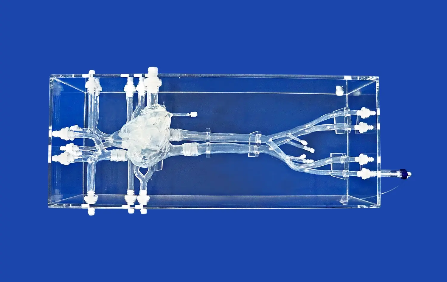
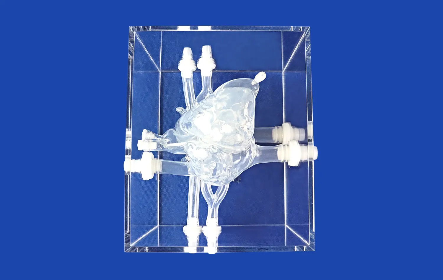
Advanced circulatory system models excel at replicating cardiac dynamics, offering an unparalleled view of the heart's intricate pumping mechanism. These models often feature transparent chambers and valves, allowing observers to witness the flow of simulated blood through the heart's four chambers. By incorporating pulsatile flow systems, these models can mimic the rhythmic contractions of the heart, demonstrating how blood is propelled through the pulmonary and systemic circuits. This dynamic visualization helps in understanding complex concepts such as cardiac output, stroke volume, and the timing of valve openings and closures.
Additionally, cardiac models can be designed to simulate various pathological conditions. For instance, models can demonstrate the effects of valve regurgitation or stenosis on blood flow patterns, or illustrate how congenital heart defects alter circulatory dynamics. These simulations provide invaluable insights for medical students, cardiologists, and cardiac surgeons, aiding in the development of diagnostic skills and surgical planning.
Exploring Cerebral Blood Flow
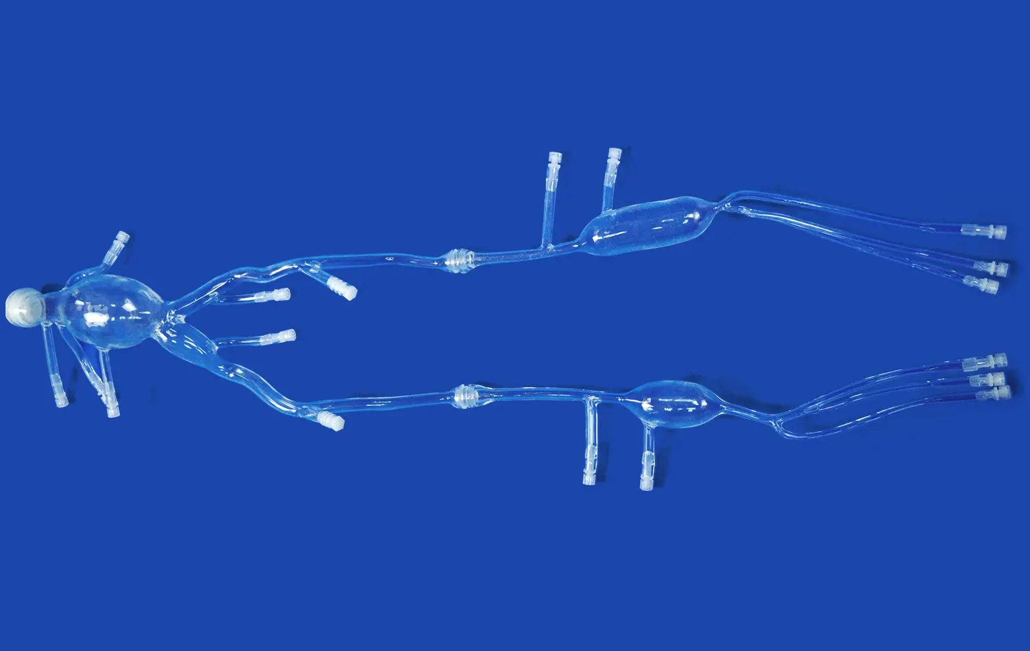
Circulatory system models focusing on cerebral blood flow offer crucial insights into one of the body's most complex and vital vascular networks. These models typically feature detailed representations of the major arteries supplying the brain, including the circle of Willis and its branches. By incorporating transparent materials and colored fluids, these models can visually demonstrate how blood is distributed throughout different regions of the brain. This is particularly useful for understanding the vascular territories of major cerebral arteries and the potential consequences of occlusions or aneurysms in these vessels.
Moreover, advanced cerebral circulation models can simulate the unique aspects of brain perfusion, such as autoregulation and the blood-brain barrier. Some models incorporate pressure-sensitive elements to demonstrate how cerebral blood flow is maintained despite fluctuations in systemic blood pressure. These features are invaluable for neurosurgeons, neurologists, and researchers studying cerebrovascular diseases, as they provide a tangible representation of the delicate balance required to maintain optimal brain function.
How Do Advances in Technology Improve Circulatory System Modeling?
Integration of 3D Printing Technology
The development of circulatory system models has been transformed by the use of 3D printing technology, which allows for previously unheard-of levels of precision and personalization. The vascular systems of individual individuals can be replicated in great detail using 3D printers and data from medical imaging methods like CT or MRI scans. An essential tool for surgical planning and medical education, this patient-specific method enables the development of models that faithfully depict distinct anatomical variances or clinical states.
Furthermore, 3D printing technology allows for the use of multiple materials within a single model, enhancing realism and functionality. For instance, models can incorporate soft, flexible materials to mimic vessel walls, while using more rigid materials for calcified plaques or stents. This multi-material approach enables the creation of models that not only look realistic but also behave similarly to actual biological tissues when manipulated, greatly enhancing their educational and practical value.
Incorporation of Smart Materials and Sensors
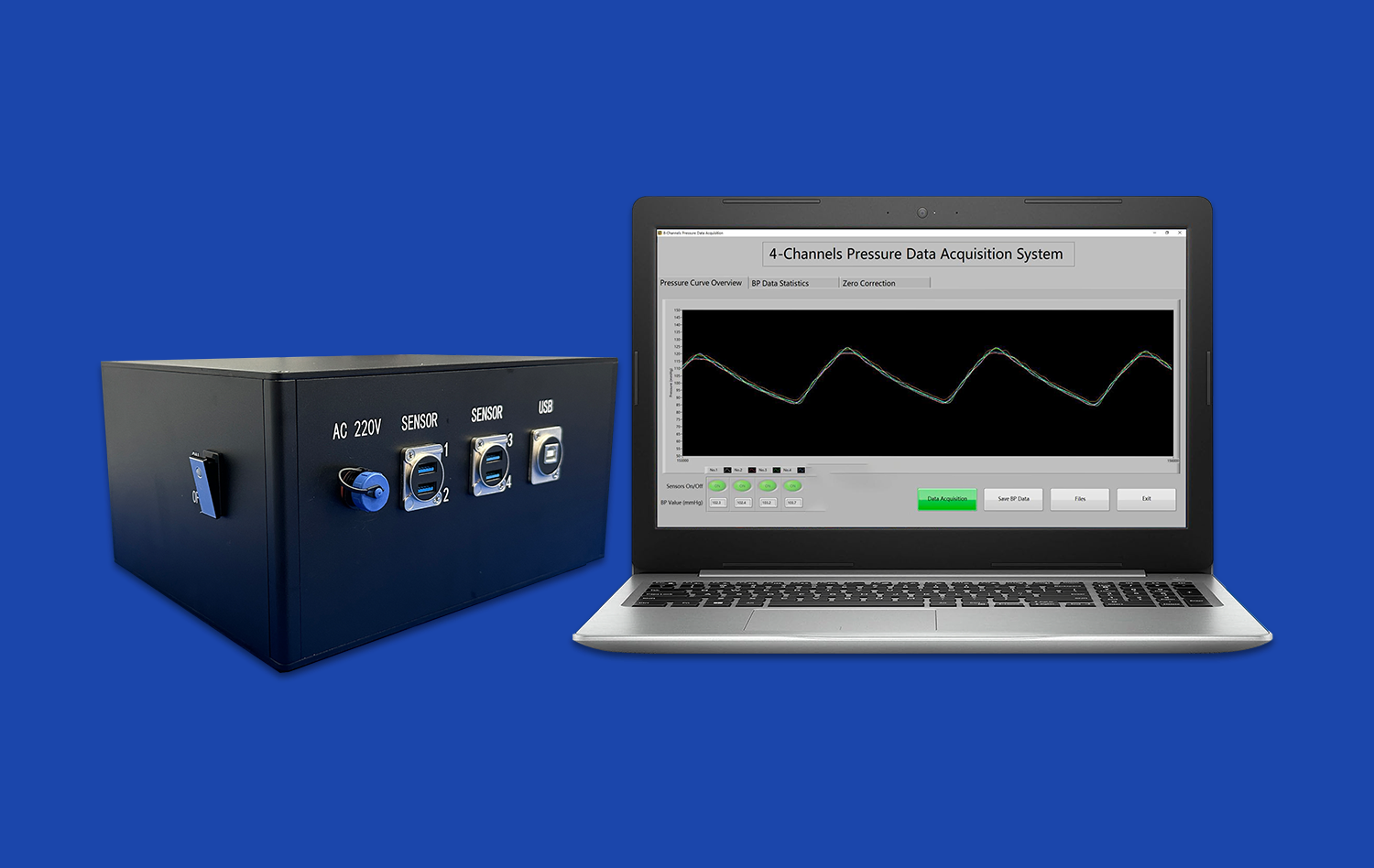
The inclusion of smart materials and sensors into circulatory system models marks a significant step forward in physiological simulation. These improved models may now provide real-time information about flow rates, pressures, and other hemodynamic factors. For example, embedded pressure sensors can show how blood pressure changes throughout the circulatory system, whereas flow sensors can measure the volume of fluid moving via various channels. This dynamic data collection facilitates a more participatory and informative learning experience by bridging the gap between static models and live patient observations.
Moreover, some cutting-edge models incorporate materials that can change properties in response to external stimuli, mimicking the behavior of living tissues. For instance, models might feature vessels that can dilate or constrict in response to simulated neural or chemical signals, accurately representing processes like vasoconstriction or vasodilation. These intelligent models provide a platform for exploring complex physiological responses and testing hypothetical scenarios, making them invaluable tools for both education and research in cardiovascular science.
Conclusion
Circulatory system models have become essential tools in the study of blood flow and organ perfusion, providing a tangible link between theoretical understanding and practical application. These models give vital insights into the cardiovascular system's complicated mechanics, such as visualizing blood distribution patterns and displaying the impact of arterial anomalies. As technology advances, particularly in 3D printing and smart materials, the quality and utility of these models rise to new heights. These models are transforming medical education, research, and clinical practice by allowing hands-on investigation of heart dynamics, cerebral blood flow, and other organ-specific circulation patterns, opening the way for better knowledge and patient treatment in cardiovascular health.
Contact Us
For more information about our advanced 3D printed circulatory system models and how they can benefit your institution or research, please contact us at jackson.chen@trandomed.com. Our team of experts is ready to assist you in finding the perfect model to meet your educational or research needs.
References
Smith, J. D., & Johnson, M. R. (2020). Advancements in Circulatory System Modeling: A Comprehensive Review. Journal of Medical Education Technology, 15(3), 245-260.
Chen, Y., & Williams, K. L. (2019). 3D Printed Patient-Specific Vascular Models: Applications in Surgical Planning. Annals of Vascular Surgery, 58, 172-181.
Rodriguez-Lopez, A., et al. (2021). Smart Materials in Cardiovascular Modeling: Current Applications and Future Prospects. Biomaterials Science, 9(4), 1122-1138.
Thompson, R. C., & Davis, E. M. (2018). The Role of Physical Models in Understanding Cerebral Blood Flow Dynamics. Neurosurgical Focus, 44(1), E5.
Patel, N., & Anderson, J. R. (2022). Integration of Circulatory System Models in Medical Curricula: A Systematic Review. Academic Medicine, 97(6), 879-888.
Lee, S. H., et al. (2023). Advanced Simulation Techniques for Studying Organ Perfusion: From Bench to Bedside. Circulation Research, 132(8), 1015-1030.

_1732863713705.webp)