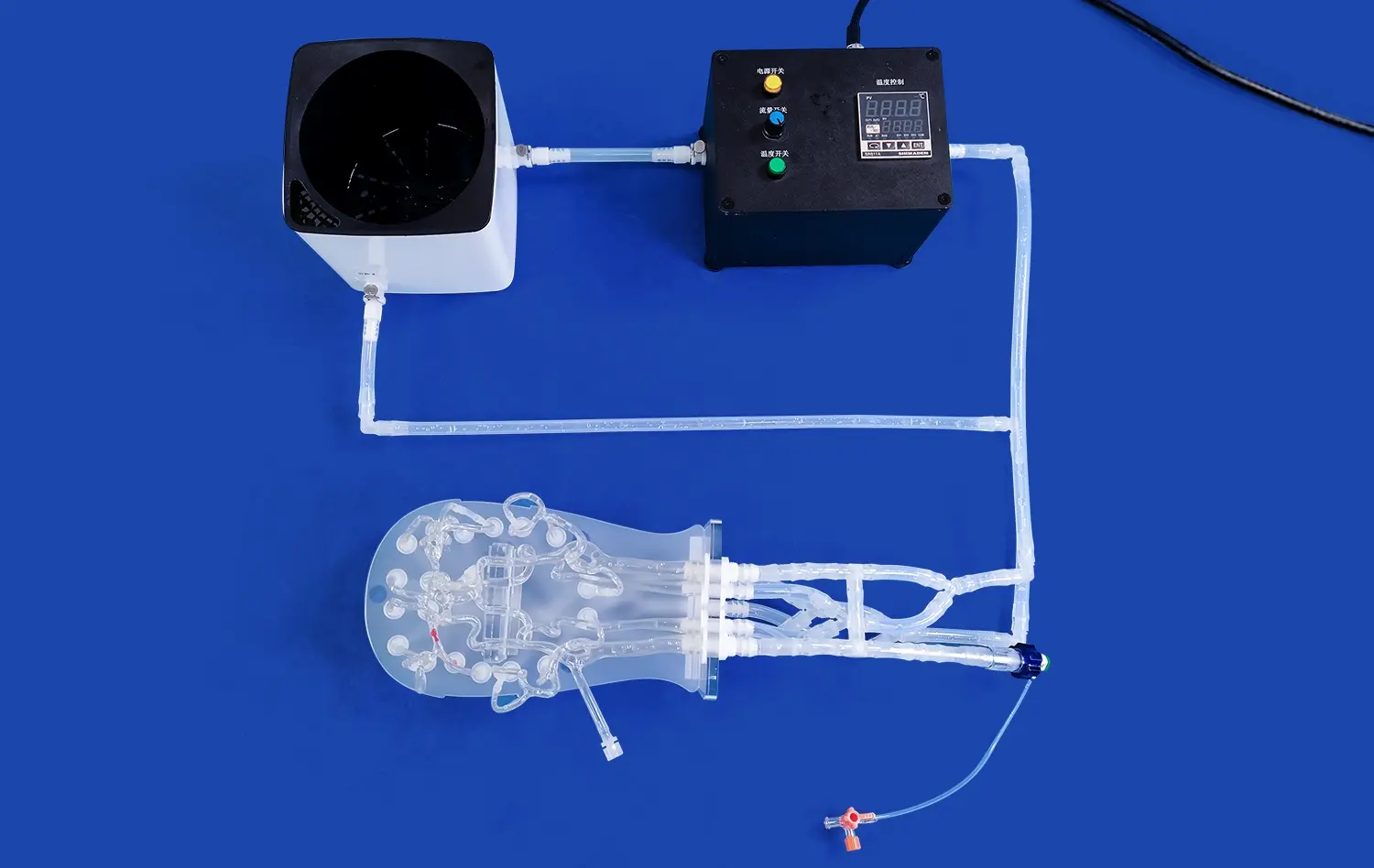
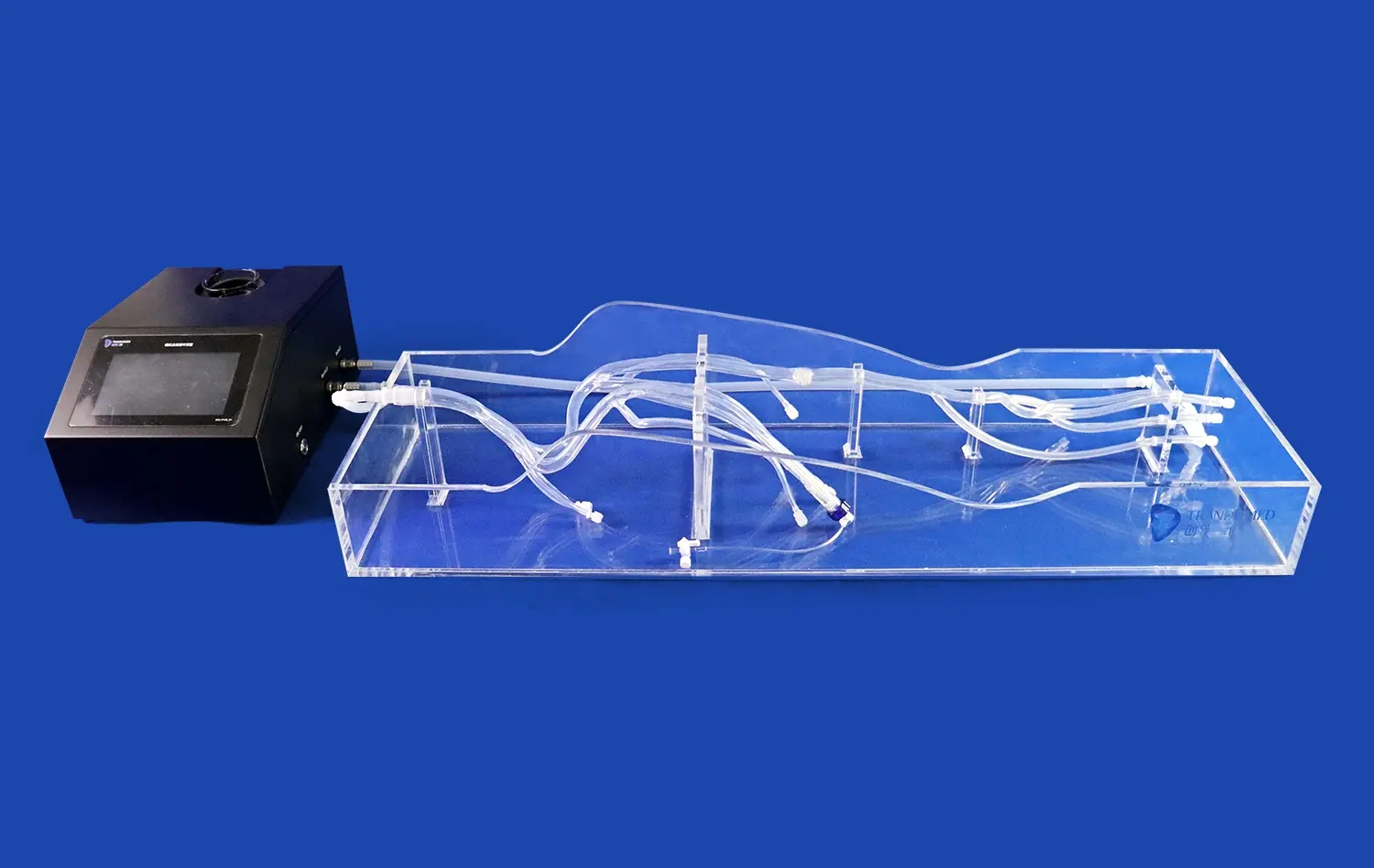
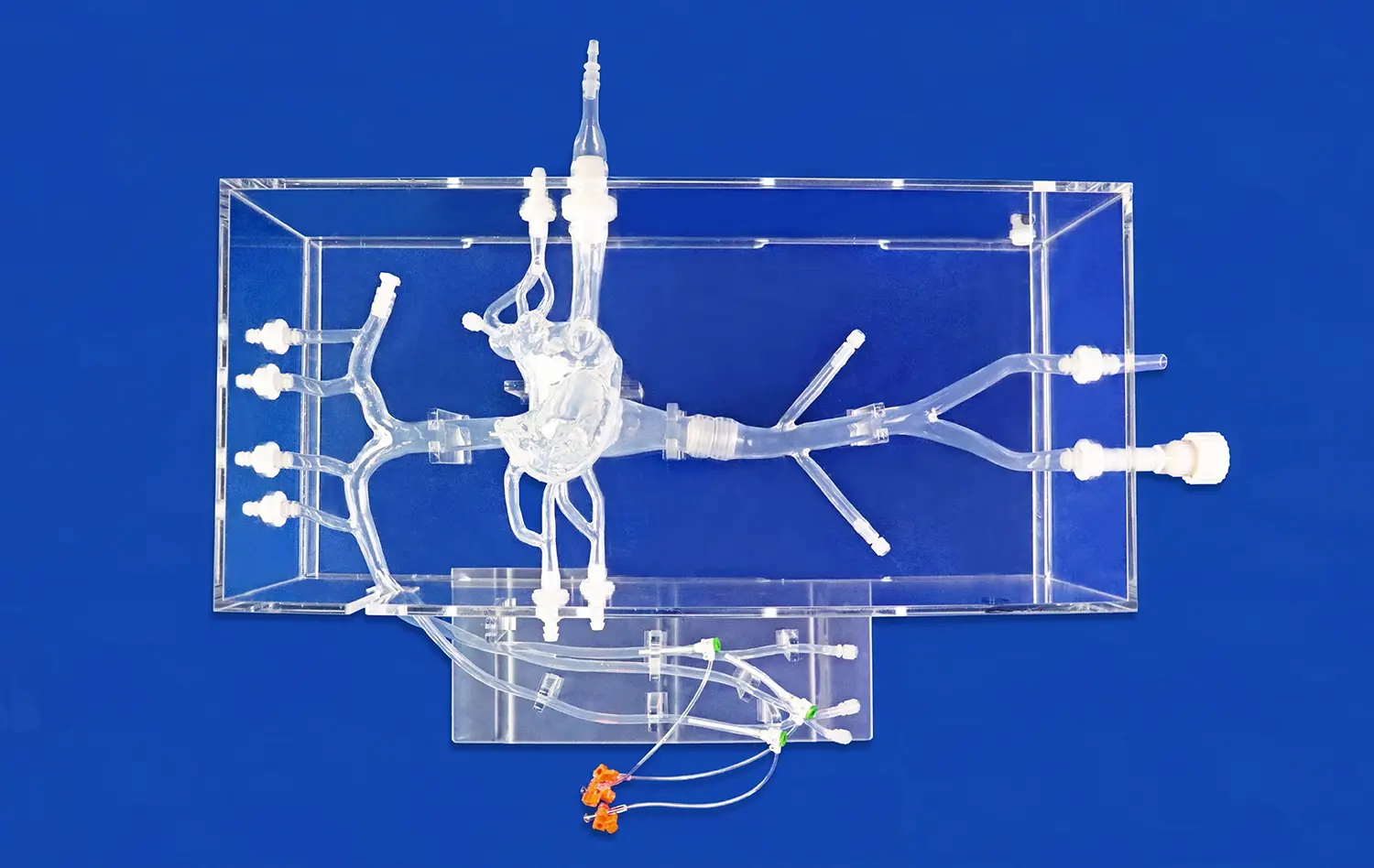
Aorta 3D models have revolutionized the approach to treating aneurysms and dissections, offering unprecedented insights into these complex cardiovascular conditions. These advanced simulations provide clinicians with detailed, patient-specific representations of aortic structures, enabling more accurate diagnoses and tailored treatment strategies. By leveraging cutting-edge 3D printing technology, medical professionals can now visualize, plan, and execute interventions with enhanced precision. The incorporation of aortic 3D models in clinical practice has significantly improved preoperative planning, intraoperative guidance, and postoperative monitoring, leading to better patient outcomes and reduced procedural risks. This innovative approach bridges the gap between imaging data and tangible, manipulable replicas, empowering healthcare providers to make more informed decisions in the management of aortic pathologies.
")
What Is the Role of Aorta 3D Models in Diagnosing Aneurysms and Dissections?
Enhanced Visualization and Spatial Understanding
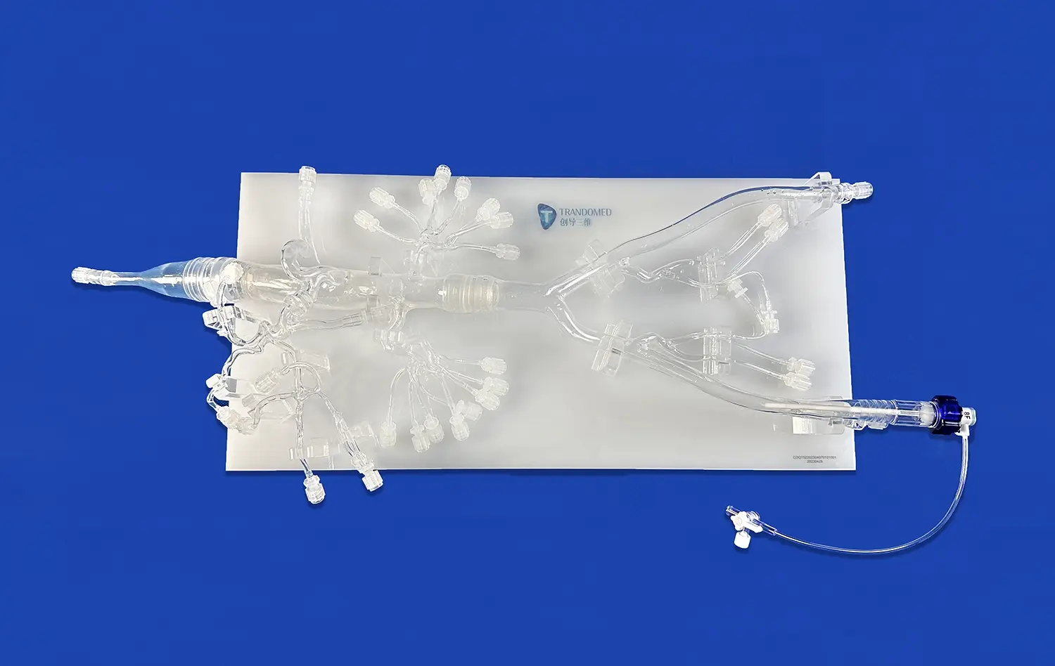
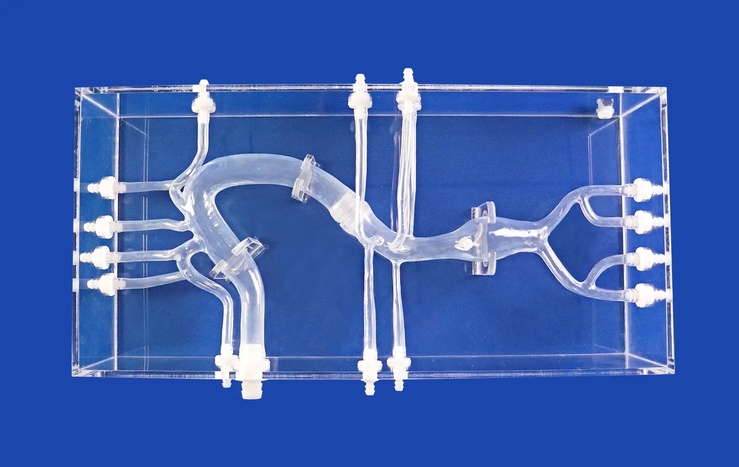
Aorta 3D models play a pivotal role in the diagnosis of aneurysms and dissections by providing unparalleled visualization of the affected vessels. Unlike traditional imaging methods, these models offer a tangible, three-dimensional representation of the aorta, allowing clinicians to examine the structure from multiple angles and perspectives. This enhanced spatial understanding is particularly valuable when dealing with complex anatomical variations or when standard imaging techniques yield ambiguous results.
The ability to manipulate and interact with a physical model of the patient's aorta enables healthcare professionals to identify subtle abnormalities that might be overlooked in 2D images. For instance, the precise location, size, and extent of an aneurysm can be more accurately determined, facilitating early detection and intervention. Similarly, the intricate patterns of aortic dissections, including the true and false lumens, entry and re-entry tears, and branch vessel involvement, become more apparent when examined in a 3D context.
Improved Communication and Patient Education
Beyond their diagnostic utility, aorta 3D models serve as powerful tools for communication among medical teams and patient education. These tangible representations bridge the gap between complex medical imaging and patient understanding, allowing for more effective explanations of the condition and proposed treatment options. Patients can visualize their own anatomy, grasp the severity of their condition, and better comprehend the rationale behind recommended interventions.
This improved communication fosters shared decision-making between healthcare providers and patients, leading to increased treatment adherence and better overall outcomes. Additionally, 3D models facilitate interdisciplinary discussions among surgeons, radiologists, and other specialists, ensuring a comprehensive approach to patient care and treatment planning.
How Do Aorta 3D Models Enhance Preoperative Planning for Aneurysm and Dissection Repair?
Customized Surgical Strategies
Aorta 3D models have transformed preoperative planning for aneurysm and dissection repair by enabling the development of customized surgical strategies. These patient-specific models allow surgeons to simulate various approaches and techniques before entering the operating room, optimizing the procedural plan for each individual case. By interacting with an accurate replica of the patient's aorta, clinicians can anticipate potential challenges, select the most appropriate surgical instruments, and determine the optimal placement of grafts or stents.
This level of precision in preoperative planning is particularly valuable in complex cases, such as thoracoabdominal aortic aneurysms or dissections involving multiple branch vessels. Surgeons can evaluate different repair options, such as open surgery, endovascular repair, or hybrid procedures, and choose the approach that offers the best balance of efficacy and safety for the patient. The ability to rehearse complicated maneuvers on a 3D model also contributes to reduced operative times and decreased risk of intraoperative complications.
Device Selection and Customization
Another significant advantage of using aorta 3D models in preoperative planning is the improved accuracy in device selection and customization. For endovascular procedures, such as thoracic endovascular aortic repair (TEVAR) or fenestrated endovascular aortic repair (FEVAR), the precise measurements obtained from 3D models ensure the selection of appropriately sized stent grafts. This reduces the risk of endoleaks, graft migration, and other complications associated with ill-fitting devices.
In cases requiring custom-made endografts, 3D models provide invaluable information for designing and manufacturing patient-specific devices. The exact geometry of the aorta, including the location and orientation of branch vessels, can be accurately replicated, leading to better-fitting and more effective implants. This level of customization not only improves the technical success of the procedure but also contributes to long-term durability and patient outcomes.
How Do Aorta 3D Models Assist in Monitoring Aneurysms and Dissections Over Time?
Tracking Disease Progression
Aorta 3D models play a crucial role in the long-term monitoring of aneurysms and dissections by providing a reliable means of tracking disease progression. By creating sequential models over time, clinicians can observe changes in the size, shape, and morphology of the affected aortic segments with unprecedented accuracy. This detailed tracking allows for the early detection of aneurysm growth or dissection progression, enabling timely intervention before complications arise.
The ability to compare aorta 3D models from different time points offers a more comprehensive understanding of disease dynamics compared to traditional imaging methods. Subtle changes in vessel wall thickness, lumen diameter, or the extent of dissection can be quantified and analyzed, providing valuable insights into the natural history of these conditions. This information not only guides treatment decisions but also contributes to our broader understanding of aortic pathologies and their progression patterns.
Evaluating Treatment Efficacy
In addition to monitoring disease progression, aorta 3D models are instrumental in evaluating the efficacy of various treatments for aneurysms and dissections. Following surgical or endovascular interventions, these models can be used to assess the success of the repair and identify any potential complications. For instance, after endovascular stent graft placement, 3D models can reveal the presence of endoleaks, graft migration, or changes in aortic morphology that might compromise the long-term success of the procedure.
The use of 3D models in post-treatment monitoring also facilitates the early detection of late complications, such as aneurysm sac expansion despite apparent successful exclusion or the development of new dissections in previously unaffected segments. This proactive approach to follow-up care allows for timely reintervention when necessary, potentially preventing catastrophic events and improving overall patient outcomes. Moreover, the insights gained from long-term monitoring using 3D models contribute to the refinement of treatment strategies and the development of more effective interventions for aortic pathologies.
Conclusion
Aorta 3D models have emerged as indispensable tools in the management of aneurysms and dissections, revolutionizing diagnosis, treatment planning, and long-term monitoring. By providing enhanced visualization, facilitating customized surgical strategies, and enabling precise tracking of disease progression, these models have significantly improved patient care and outcomes. As technology continues to advance, the integration of aorta 3D models into clinical practice is expected to expand further, offering new possibilities for personalized medicine in the field of vascular surgery. The continued development and refinement of these models promise to drive innovation in aortic disease management, ultimately leading to more effective treatments and better quality of life for patients affected by these complex cardiovascular conditions.
Contact Us
To learn more about our advanced aorta 3D models and how they can enhance your clinical practice, please contact us at jackson.chen@trandomed.com. Our team of experts is ready to assist you in leveraging this cutting-edge technology for improved patient care.
References
Wang, H., et al. (2020). "Application of 3D printing technology in the treatment of aortic dissection." Journal of Thoracic Disease, 12(3), 1044-1052.
Tam, C. H., et al. (2018). "Use of 3-dimensional printed models in planning the treatment of aortic aneurysms." Journal of Vascular Surgery, 68(3), 903-911.
Elefteriades, J. A., et al. (2019). "3D-printed aortic models for surgical planning in complex aortic aneurysm cases." Annals of Thoracic Surgery, 107(5), 1478-1483.
Sun, Z., et al. (2017). "3D printing in cardiovascular disease: Current applications and future challenges." Quantitative Imaging in Medicine and Surgery, 7(3), 380-391.
Giannopoulos, A. A., et al. (2016). "Applications of 3D printing in cardiovascular diseases." Nature Reviews Cardiology, 13(12), 701-718.
Mahmood, F., et al. (2015). "Three-dimensional printing of mitral valve using echocardiographic data." JACC: Cardiovascular Imaging, 8(2), 227-229.
_1732863962417.webp)