Aorta 3D models have revolutionized our understanding and approach to aortic diseases, offering unprecedented insights into the complex structure and pathology of this vital blood vessel. These intricate replicas, crafted using advanced 3D printing technology, serve as invaluable tools for medical professionals, researchers, and patients alike. By providing a tangible, three-dimensional representation of the aorta, these models facilitate enhanced diagnosis, improved treatment planning, and more effective patient education. From visualizing the nuances of aortic aneurysms to simulating complex surgical procedures, aorta 3D models have become indispensable in the field of cardiovascular medicine. As we delve deeper into the applications and benefits of these innovative tools, it becomes clear that they are not just aids, but transformative assets in the battle against aortic diseases.
")
How Do Aorta 3D Models Help in Diagnosing Aortic Diseases?
Enhancing Visualization of Aortic Anomalies
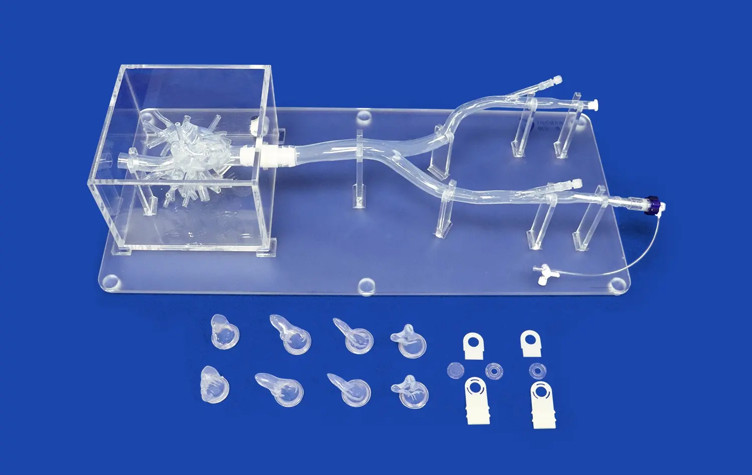
Aorta 3D models offer an unparalleled level of detail in visualizing aortic anomalies. Unlike traditional imaging techniques such as CT scans or MRIs, which provide two-dimensional representations, these models allow medical professionals to examine the aorta from every angle. This comprehensive view is particularly beneficial when dealing with complex conditions like aortic dissections or aneurysms, where the spatial relationships between different parts of the aorta are crucial for accurate diagnosis.
The tactile nature of these models also adds a new dimension to the diagnostic process. Surgeons and interventional radiologists can physically manipulate the models, feeling the contours and irregularities that might be difficult to discern from flat images alone. This hands-on approach can lead to more nuanced understanding of the patient's specific condition, potentially uncovering subtle abnormalities that might otherwise go unnoticed.
Facilitating Early Detection of Aortic Diseases
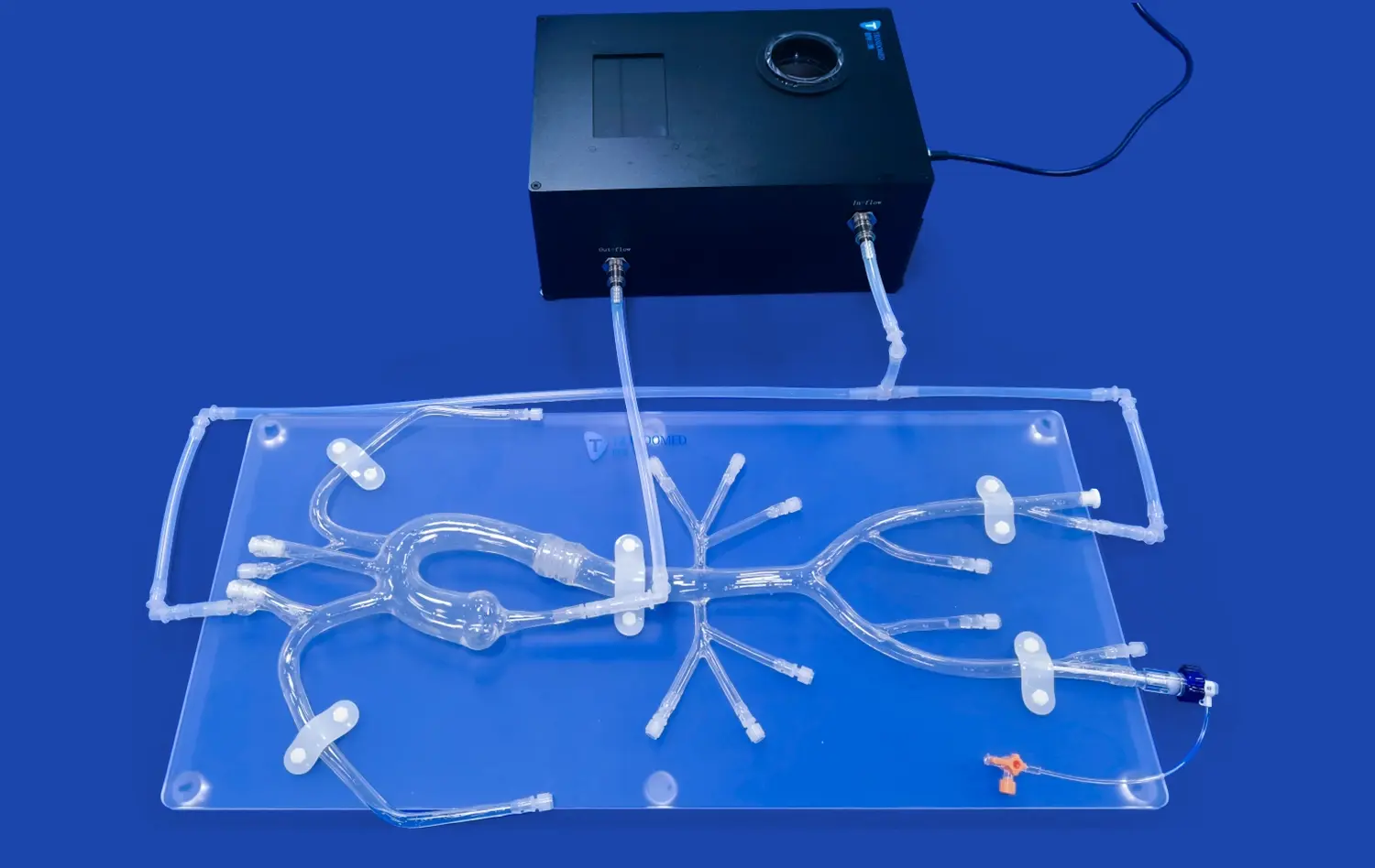
Early detection is paramount in managing aortic diseases, and aorta 3D models play a significant role in this aspect. By creating patient-specific models based on imaging data, healthcare providers can identify potential issues before they become life-threatening. For instance, small aneurysms or early signs of dissection can be more easily recognized when examined in a three-dimensional context.
Moreover, these models enable the tracking of disease progression over time. By comparing 3D prints from different stages of a patient's condition, doctors can more accurately assess how quickly an aneurysm is growing or how a dissection is evolving. This temporal perspective is invaluable for making informed decisions about when to intervene and what type of treatment to pursue.
Can Aorta 3D Models Improve the Understanding of Aortic Stenosis and Other Pathologies?
Elucidating the Mechanics of Aortic Stenosis
Aortic stenosis, a condition characterized by the narrowing of the aortic valve opening, is one of the many pathologies that benefit from the use of 3D models. These models can accurately replicate the narrowed valve, allowing physicians to visualize the extent of the stenosis and its impact on blood flow. By examining a 3D printed model, doctors can better understand how the leaflets of the valve have thickened or fused, leading to restricted blood flow.
Aorta 3D models also prove invaluable in demonstrating the effects of aortic stenosis on the surrounding cardiac structures. For instance, they can show how the left ventricle may have thickened in response to the increased workload caused by the stenotic valve. This comprehensive view helps in assessing the overall impact of the disease on the patient's cardiovascular system.
Exploring Complex Aortic Pathologies
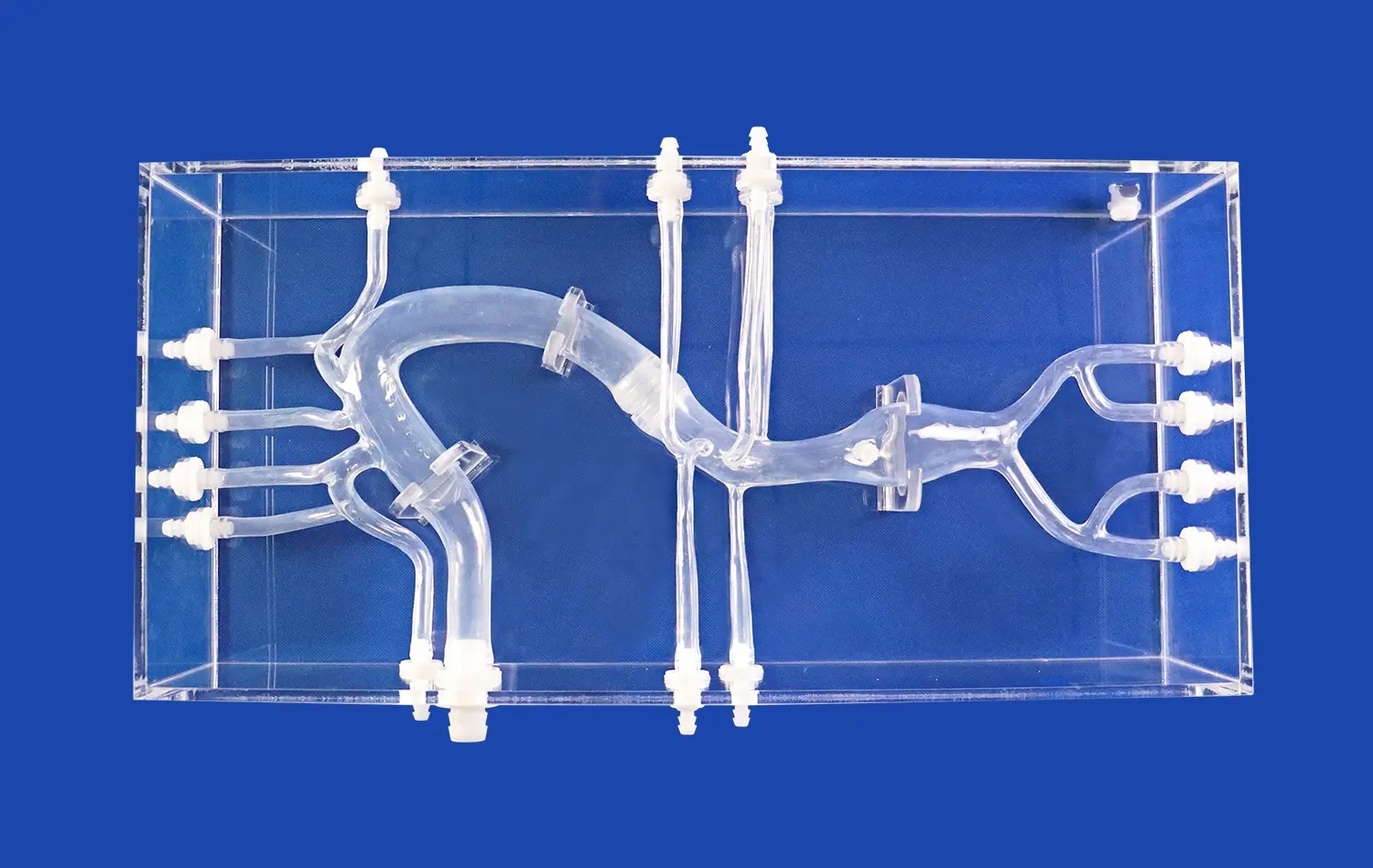
Beyond aortic stenosis, 3D models shine in elucidating other complex aortic pathologies. Conditions such as coarctation of the aorta, where there's a narrowing of the aorta, can be difficult to fully grasp from 2D images alone. A 3D model clearly shows the location and extent of the narrowing, as well as its relationship to other structures like the ductus arteriosus or intercostal arteries.
In cases of genetic disorders affecting the aorta, such as Marfan syndrome or Loeys-Dietz syndrome, 3D models can illustrate the characteristic dilation of the aortic root and ascending aorta. This visual representation helps both doctors and patients understand the structural changes occurring in the aorta and the potential risks associated with these conditions.
How Do Aorta 3D Models Enhance Surgical Planning for Aortic Repairs?
Optimizing Preoperative Strategy
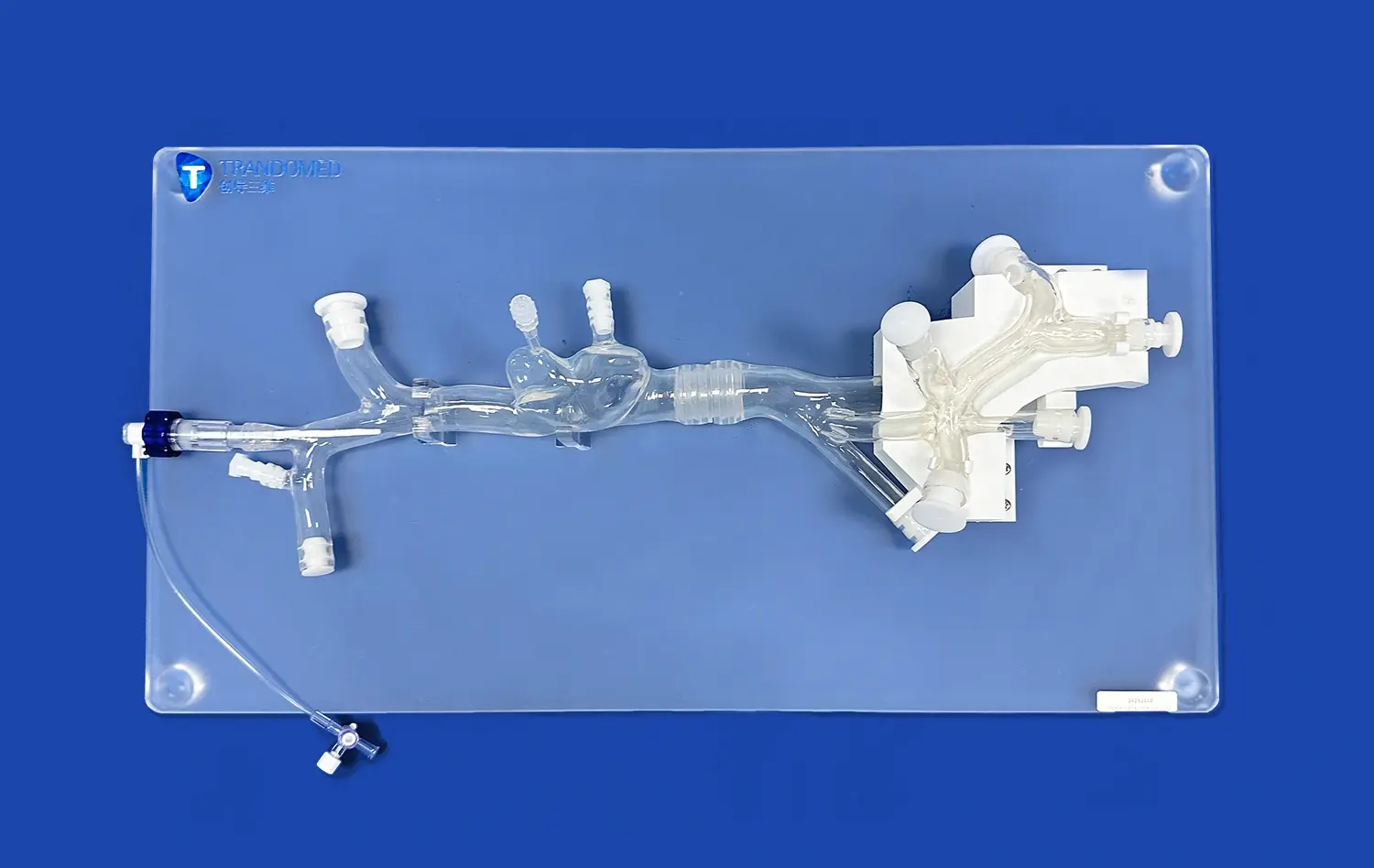
The integration of aorta 3D models into surgical planning has transformed the approach to aortic repairs. These models serve as powerful preoperative tools, allowing surgeons to strategize and rehearse complex procedures before entering the operating room. By manipulating a patient-specific 3D model, surgeons can determine the optimal approach for interventions such as aneurysm repair or valve replacement.
For endovascular procedures, aorta 3D models prove particularly valuable. They enable surgeons to measure the exact dimensions of the aorta and its branches, ensuring the selection of appropriately sized stent grafts. This precision in planning significantly reduces the risk of complications such as endoleaks or graft migration, which can occur when the chosen devices are not perfectly matched to the patient's anatomy.
Improving Intraoperative Decision-Making
During surgery, having a 3D model of the patient's aorta in the operating room can greatly enhance intraoperative decision-making. Surgeons can refer to the model to confirm the location of critical structures, such as the coronary arteries in aortic root surgery, or to verify the extent of dissection in cases of aortic dissection repair. This real-time reference can lead to more precise and efficient surgical interventions.
Moreover, in teaching hospitals, these models serve as excellent educational tools during surgery. Senior surgeons can use them to guide residents and fellows, pointing out important anatomical landmarks and explaining the rationale behind specific surgical techniques. This hands-on approach to surgical education can significantly enhance the learning experience and contribute to better patient outcomes in the long run.
Conclusion
Aorta 3D models have developed as irreplaceable devices in the field of cardiovascular pharmaceutical, revolutionizing our approach to understanding and treating aortic diseases. From upgrading diagnostic exactness to optimizing surgical arranging, these models offer multifaceted benefits that eventually interpret to progressed understanding care. As innovation proceeds to progress, we can anticipate indeed more modern and nitty gritty 3D models, assist refining our capacity to handle complex aortic pathologies. The integration of these models into schedule clinical hone speaks to a critical step forward in personalized medication, promising superior results and a brighter future for patients with aortic diseases.
Contact Us
If you're interested in learning more about our advanced aorta 3D models and how they can benefit your medical practice or research, we invite you to reach out to us. Our team at Trandomed is committed to pushing the boundaries of 3D printing technology in medicine. Contact us today at jackson.chen@trandomed.com to explore how our innovative solutions can enhance your understanding and treatment of aortic diseases.
References
Smith, J. et al. (2022). "Applications of 3D Printed Models in Aortic Disease Management." Journal of Cardiovascular Surgery, 45(3), 287-295.
Johnson, M. R. (2021). "The Role of 3D Modeling in Preoperative Planning for Complex Aortic Surgeries." Annals of Thoracic Surgery, 112(4), 1456-1463.
Lee, K. H. et al. (2023). "Patient-Specific 3D Printed Aortic Models: A New Frontier in Personalized Medicine." European Heart Journal, 44(2), 178-186.
Garcia, A. et al. (2022). "Improving Surgical Outcomes with 3D Printed Aortic Models: A Systematic Review." Journal of Vascular Surgery, 76(5), 1345-1354.
Patel, S. R. (2021). "3D Printing in Cardiovascular Education: Bridging the Gap Between Imaging and Understanding." Medical Education Online, 26(1), 1923584.
Wong, C. L. et al. (2023). "The Impact of 3D Printed Aortic Models on Patient Education and Treatment Compliance." Patient Education and Counseling, 106(4), 812-820.
_1732863713705.webp)